- PDB-8j0l: Structure of DNA binding Domain of Human TFAP2A -
+
Open data
ID or keywords:
Loading...
-
Basic information
Entry
Database: PDB / ID: 8j0l
Title
Structure of DNA binding Domain of Human TFAP2A
Components
Transcription factor AP-2-alpha
Keywords
DNA BINDING PROTEIN / TFAP2 family
Function / homology
Function and homology information
optic vesicle morphogenesis / optic cup structural organization / oculomotor nerve formation / positive regulation of tooth mineralization / trigeminal nerve development / TFAP2 (AP-2) family regulates transcription of other transcription factors / Positive Regulation of CDH1 Gene Transcription / TFAP2 (AP-2) family regulates transcription of cell cycle factors / TFAP2A acts as a transcriptional repressor during retinoic acid induced cell differentiation / Specification of the neural plate border ...optic vesicle morphogenesis / optic cup structural organization / oculomotor nerve formation / positive regulation of tooth mineralization / trigeminal nerve development / TFAP2 (AP-2) family regulates transcription of other transcription factors / Positive Regulation of CDH1 Gene Transcription / TFAP2 (AP-2) family regulates transcription of cell cycle factors / TFAP2A acts as a transcriptional repressor during retinoic acid induced cell differentiation / Specification of the neural plate border / Negative regulation of activity of TFAP2 (AP-2) family transcription factors / retina layer formation / Transcriptional regulation by the AP-2 (TFAP2) family of transcription factors / : / Developmental Lineage of Mammary Stem Cells / embryonic cranial skeleton morphogenesis / Activation of the TFAP2 (AP-2) family of transcription factors / embryonic forelimb morphogenesis / bone morphogenesis / inner ear morphogenesis / eyelid development in camera-type eye / SUMOylation of transcription factors / roof of mouth development / Regulation of MITF-M-dependent genes involved in pigmentation / negative regulation of reactive oxygen species metabolic process / regulation of cell differentiation / positive regulation of bone mineralization / TFAP2 (AP-2) family regulates transcription of growth factors and their receptors / RNA polymerase II transcription regulatory region sequence-specific DNA binding / skeletal system development / kidney development / cellular response to iron ion / sensory perception of sound / DNA-binding transcription repressor activity, RNA polymerase II-specific / sequence-specific double-stranded DNA binding / positive regulation of neuron apoptotic process / nervous system development / regulation of cell population proliferation / DNA-binding transcription activator activity, RNA polymerase II-specific / sequence-specific DNA binding / DNA-binding transcription factor activity, RNA polymerase II-specific / transcription cis-regulatory region binding / RNA polymerase II cis-regulatory region sequence-specific DNA binding / negative regulation of cell population proliferation / negative regulation of DNA-templated transcription / chromatin binding / positive regulation of gene expression / negative regulation of apoptotic process / positive regulation of DNA-templated transcription / chromatin / DNA-templated transcription / negative regulation of transcription by RNA polymerase II / positive regulation of transcription by RNA polymerase II / DNA binding / nucleoplasm / identical protein binding / nucleus Similarity search - Function
 Movie
Movie Controller


 Open data
 Basic information
 Components
 Keywords
 Function and homology information
 Homo sapiens (human)
 X-RAY DIFFRACTION /
  Authors
 China, 1items
  Citation
 Structure visualization
 Downloads & links
 Other downloads
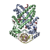


 PDBj
PDBj


 Assembly
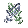


 Mass: 92.094 Da / Num. of mol.: 2 / Source method: obtained synthetically / Formula: C3H8O3
 Mass: 18.015 Da / Num. of mol.: 192 / Source method: isolated from a natural source / Formula: H2O
 Sample preparation
 Processing