 ムービー
ムービー コントローラー


+ データを開く

- 基本情報
基本情報
| 登録情報 | データベース: PDB / ID: 8izu | ||||||
|---|---|---|---|---|---|---|---|
| タイトル | Crystal structure of the N-terminal domain (residues 1-137) of MPXV A7 | ||||||
 要素 要素 | Virion morphogenesis protein OPG132 | ||||||
 キーワード キーワード | VIRAL PROTEIN / MPXV / A7 / N-terminus | ||||||
| 機能・相同性 | Poxvirus A6 / Poxvirus A6 protein / virion component / host cell cytoplasm / NICKEL (II) ION / Virion morphogenesis protein OPG132 機能・相同性情報 機能・相同性情報 | ||||||
| 生物種 |  Monkeypox virus (サル痘ウイルス) Monkeypox virus (サル痘ウイルス) | ||||||
| 手法 |  X線回折 / シンクロトロン / 分子置換 / 解像度: 2.54 Å X線回折 / シンクロトロン / 分子置換 / 解像度: 2.54 Å | ||||||
 データ登録者 データ登録者 | Ni, X.C. / Lei, J. | ||||||
| 資金援助 |  中国, 1件 中国, 1件
| ||||||
 引用 引用 | ジャーナル: Virol Sin / 年: 2024 タイトル: Structural analysis of conformational changes in the mpox virus A7 protein. 著者: Ni, X. / Wang, K. / Han, Y. / Lei, J. | ||||||
| 履歴 |
|
- 構造の表示
構造の表示
| 構造ビューア | 分子: MolmilJmol/JSmol |
|---|
- ダウンロードとリンク
-ダウンロード
| PDBx/mmCIF形式 | 8izu.cif.gz | 70.4 KB | 表示 | PDBx/mmCIF形式 |
|---|---|---|---|---|
| PDB形式 | pdb8izu.ent.gz | 51.5 KB | 表示 | PDB形式 |
| PDBx/mmJSON形式 | 8izu.json.gz | ツリー表示 | PDBx/mmJSON形式 | |
| その他 |  その他のダウンロード その他のダウンロード |
-検証レポート
| 文書・要旨 | 8izu_validation.pdf.gz | 443.9 KB | 表示 | wwPDB検証レポート |
|---|---|---|---|---|
| 文書・詳細版 | 8izu_full_validation.pdf.gz | 444.5 KB | 表示 | |
| XML形式データ | 8izu_validation.xml.gz | 12.7 KB | 表示 | |
| CIF形式データ | 8izu_validation.cif.gz | 16.9 KB | 表示 | |
| アーカイブディレクトリ | https://data.pdbj.org/pub/pdb/validation_reports/iz/8izuftp://data.pdbj.org/pub/pdb/validation_reports/iz/8izu | HTTPS FTP |
-関連構造データ

-リンク
 PDBj
PDBj- 集合体
集合体
| 登録構造単位 | 
| ||||||||
|---|---|---|---|---|---|---|---|---|---|
| 1 | 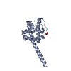
| ||||||||
| 2 | 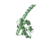
| ||||||||
| 単位格子 |
|
-要素
| #1: タンパク質 | 分子量: 16157.606 Da / 分子数: 2 / 断片: N-terminal domain(residues 1-137) / 由来タイプ: 組換発現 由来: (組換発現) Monkeypox virus (サル痘ウイルス)遺伝子: OPG132, MPXVgp117 / 発現宿主:  #2: 化合物 | ChemComp-GOL / |   分子量: 92.094 Da / 分子数: 1 / 由来タイプ: 合成 / 式: C3H8O3 分子量: 92.094 Da / 分子数: 1 / 由来タイプ: 合成 / 式: C3H8O3#3: 化合物 | ChemComp-NI / |   分子量: 58.693 Da / 分子数: 1 / 由来タイプ: 合成 / 式: Ni 分子量: 58.693 Da / 分子数: 1 / 由来タイプ: 合成 / 式: Ni#4: 水 | ChemComp-HOH / |  分子量: 18.015 Da / 分子数: 87 / 由来タイプ: 天然 / 式: H2O 分子量: 18.015 Da / 分子数: 87 / 由来タイプ: 天然 / 式: H2O研究の焦点であるリガンドがあるか | N | |
|---|
-実験情報
-実験
| 実験 | 手法: X線回折 / 使用した結晶の数: 1 |
|---|
- 試料調製
試料調製
| 結晶 | マシュー密度: 3.16 Å3/Da / 溶媒含有率: 61.12 % |
|---|---|
| 結晶化 | 温度: 291 K / 手法: 蒸気拡散法, シッティングドロップ法 詳細: 0.1 M Potassium thiocyanate, 30% w/v Polyethylene glycol monomethyl ether 2000 |
-データ収集
| 回折 | 平均測定温度: 100 K / Serial crystal experiment: N |
|---|---|
| 放射光源 | 由来: シンクロトロン / サイト: SSRF / ビームライン: BL19U1 / 波長: 0.97851 Å |
| 検出器 | タイプ: DECTRIS PILATUS 6M / 検出器: PIXEL / 日付: 2022年12月10日 |
| 放射 | プロトコル: SINGLE WAVELENGTH / 単色(M)・ラウエ(L): M / 散乱光タイプ: x-ray |
| 放射波長 | 波長: 0.97851 Å / 相対比: 1 |
| 反射 | 解像度: 2.54→19.45 Å / Num. obs: 14013 / % possible obs: 99.8 % / 冗長度: 12.8 % / CC1/2: 0.997 / Net I/σ(I): 14.1 |
| 反射 シェル | 解像度: 2.54→2.65 Å / Num. unique obs: 1676 / CC1/2: 0.954 |
- 解析
解析
| ソフトウェア |
| ||||||||||||||||||||||||||||||||||||||||||||||||||||||||||||||||||||||||||||||||||||||||||||||||||||||||||||||||||||||||||||||||||||||||||||||||||||||||||||||||||||||||||||||||||||||
|---|---|---|---|---|---|---|---|---|---|---|---|---|---|---|---|---|---|---|---|---|---|---|---|---|---|---|---|---|---|---|---|---|---|---|---|---|---|---|---|---|---|---|---|---|---|---|---|---|---|---|---|---|---|---|---|---|---|---|---|---|---|---|---|---|---|---|---|---|---|---|---|---|---|---|---|---|---|---|---|---|---|---|---|---|---|---|---|---|---|---|---|---|---|---|---|---|---|---|---|---|---|---|---|---|---|---|---|---|---|---|---|---|---|---|---|---|---|---|---|---|---|---|---|---|---|---|---|---|---|---|---|---|---|---|---|---|---|---|---|---|---|---|---|---|---|---|---|---|---|---|---|---|---|---|---|---|---|---|---|---|---|---|---|---|---|---|---|---|---|---|---|---|---|---|---|---|---|---|---|---|---|---|---|
| 精密化 | 構造決定の手法: 分子置換 開始モデル: 8IZT 解像度: 2.54→19.45 Å / Cor.coef. Fo:Fc: 0.961 / Cor.coef. Fo:Fc free: 0.937 / SU B: 11.157 / SU ML: 0.217 / 交差検証法: THROUGHOUT / ESU R: 0.399 / ESU R Free: 0.258 / 立体化学のターゲット値: MAXIMUM LIKELIHOOD / 詳細: HYDROGENS HAVE BEEN ADDED IN THE RIDING POSITIONS
| ||||||||||||||||||||||||||||||||||||||||||||||||||||||||||||||||||||||||||||||||||||||||||||||||||||||||||||||||||||||||||||||||||||||||||||||||||||||||||||||||||||||||||||||||||||||
| 溶媒の処理 | イオンプローブ半径: 0.8 Å / 減衰半径: 0.8 Å / VDWプローブ半径: 1.2 Å / 溶媒モデル: MASK | ||||||||||||||||||||||||||||||||||||||||||||||||||||||||||||||||||||||||||||||||||||||||||||||||||||||||||||||||||||||||||||||||||||||||||||||||||||||||||||||||||||||||||||||||||||||
| 原子変位パラメータ | Biso mean: 69.853 Å2
| ||||||||||||||||||||||||||||||||||||||||||||||||||||||||||||||||||||||||||||||||||||||||||||||||||||||||||||||||||||||||||||||||||||||||||||||||||||||||||||||||||||||||||||||||||||||
| 精密化ステップ | サイクル: 1 / 解像度: 2.54→19.45 Å
| ||||||||||||||||||||||||||||||||||||||||||||||||||||||||||||||||||||||||||||||||||||||||||||||||||||||||||||||||||||||||||||||||||||||||||||||||||||||||||||||||||||||||||||||||||||||
| 拘束条件 |
|