 Movie
Movie Controller


+ Open data

- Basic information
| Entry | Database: PDB / ID: 8ir0 | ||||||
|---|---|---|---|---|---|---|---|
| Title | AfFer mutant-P156F | ||||||
 Components Components | Ferritin | ||||||
 Keywords Keywords | METAL BINDING PROTEIN / Asterias forbesii ferritin / mutant-P156F | ||||||
| Function / homology |  Function and homology information Function and homology informationferroxidase / ferroxidase activity / ferric iron binding / iron ion transport / ferrous iron binding / intracellular iron ion homeostasis / cytoplasm Similarity search - Function | ||||||
| Biological species |  Asterias forbesi (Forbes's starfish) Asterias forbesi (Forbes's starfish) | ||||||
| Method |  X-RAY DIFFRACTION / SYNCHROTRON / MOLECULAR REPLACEMENT / Resolution: 2.89 Å X-RAY DIFFRACTION / SYNCHROTRON / MOLECULAR REPLACEMENT / Resolution: 2.89 Å | ||||||
 Authors Authors | Zhao, G. / Zhang, C. / Zang, J. / Zhang, T. | ||||||
| Funding support | 1items
| ||||||
 Citation Citation | Journal: Foods / Year: 2023 Title: Preparation and Unique Three-Dimensional Self-Assembly Property of Starfish Ferritin. Authors: Zhang, C. / Chen, X. / Liu, B. / Zang, J. / Zhang, T. / Zhao, G. | ||||||
| History |
|
- Structure visualization
| Structure viewer | Molecule: MolmilJmol/JSmol |
|---|
- Downloads & links
-Download
| PDBx/mmCIF format | 8ir0.cif.gz | 129.8 KB | Display | PDBx/mmCIF format |
|---|---|---|---|---|
| PDB format | pdb8ir0.ent.gz | 83.1 KB | Display | PDB format |
| PDBx/mmJSON format | 8ir0.json.gz | Tree view | PDBx/mmJSON format | |
| Others |  Other downloads Other downloads |
-Validation report
| Summary document | 8ir0_validation.pdf.gz | 429.2 KB | Display | wwPDB validaton report |
|---|---|---|---|---|
| Full document | 8ir0_full_validation.pdf.gz | 433.1 KB | Display | |
| Data in XML | 8ir0_validation.xml.gz | 18.2 KB | Display | |
| Data in CIF | 8ir0_validation.cif.gz | 24.3 KB | Display | |
| Arichive directory | https://data.pdbj.org/pub/pdb/validation_reports/ir/8ir0ftp://data.pdbj.org/pub/pdb/validation_reports/ir/8ir0 | HTTPS FTP |
-Related structure data


-Links
 PDBj
PDBj


- Assembly
| Deposited unit | 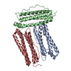
| ||||||||||||
|---|---|---|---|---|---|---|---|---|---|---|---|---|---|
| 1 | x 8
| ||||||||||||
| Unit cell |
|
-Components
| #1: Protein | Mass: 19302.516 Da / Num. of mol.: 3 / Mutation: P156F Source method: isolated from a genetically manipulated source Source: (gene. exp.) Asterias forbesi (Forbes's starfish) / Production host:  |
|---|
-Experimental details
-Experiment
| Experiment | Method: X-RAY DIFFRACTION / Number of used crystals: 1 |
|---|
- Sample preparation
| Crystal | Density Matthews: 3.03 Å3/Da / Density % sol: 59.38 % |
|---|---|
| Crystal grow | Temperature: 293 K / Method: vapor diffusion, hanging drop Details: 100 mM MES, 20%(v/v) 1,4-butanediol, 200 mM Lithium sulfate, pH 6.0 |
-Data collection
| Diffraction | Mean temperature: 100 K / Serial crystal experiment: N |
|---|---|
| Diffraction source | Source: SYNCHROTRON / Site: SSRF  / Beamline: BL18U1 / Wavelength: 0.9792 Å / Beamline: BL18U1 / Wavelength: 0.9792 Å |
| Detector | Type: DECTRIS PILATUS3 6M / Detector: PIXEL / Date: Jan 2, 2022 |
| Radiation | Protocol: SINGLE WAVELENGTH / Monochromatic (M) / Laue (L): M / Scattering type: x-ray |
| Radiation wavelength | Wavelength: 0.9792 Å / Relative weight: 1 |
| Reflection | Resolution: 2.89→29.65 Å / Num. obs: 16300 / % possible obs: 99.71 % / Redundancy: 12.7 % / Biso Wilson estimate: 45.11 Å2 / CC1/2: 0.972 / Net I/σ(I): 2 |
| Reflection shell | Resolution: 2.9→2.95 Å / Rmerge(I) obs: 0.519 / Num. unique obs: 16300 |
- Processing
| Software |
| |||||||||||||||||||||||||||||||||||||||||||||||||||||||||||||||||||||||||||||||||||||||||||
|---|---|---|---|---|---|---|---|---|---|---|---|---|---|---|---|---|---|---|---|---|---|---|---|---|---|---|---|---|---|---|---|---|---|---|---|---|---|---|---|---|---|---|---|---|---|---|---|---|---|---|---|---|---|---|---|---|---|---|---|---|---|---|---|---|---|---|---|---|---|---|---|---|---|---|---|---|---|---|---|---|---|---|---|---|---|---|---|---|---|---|---|---|
| Refinement | Method to determine structure: MOLECULAR REPLACEMENT / Resolution: 2.89→29.65 Å / SU ML: 0.2952 / Cross valid method: FREE R-VALUE / σ(F): 1.36 / Phase error: 23.0965 Stereochemistry target values: GeoStd + Monomer Library + CDL v1.2
| |||||||||||||||||||||||||||||||||||||||||||||||||||||||||||||||||||||||||||||||||||||||||||
| Solvent computation | Shrinkage radii: 0.9 Å / VDW probe radii: 1.11 Å / Solvent model: FLAT BULK SOLVENT MODEL | |||||||||||||||||||||||||||||||||||||||||||||||||||||||||||||||||||||||||||||||||||||||||||
| Displacement parameters | Biso mean: 38.63 Å2 | |||||||||||||||||||||||||||||||||||||||||||||||||||||||||||||||||||||||||||||||||||||||||||
| Refinement step | Cycle: LAST / Resolution: 2.89→29.65 Å
| |||||||||||||||||||||||||||||||||||||||||||||||||||||||||||||||||||||||||||||||||||||||||||
| Refine LS restraints |
| |||||||||||||||||||||||||||||||||||||||||||||||||||||||||||||||||||||||||||||||||||||||||||
| LS refinement shell |
|
