 Movie
Movie Controller

[English] 日本語
 Yorodumi
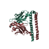
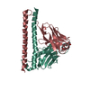
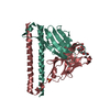
- PDB-8iqr: Crystal structure of Anti-PEG antibody M9 Fv-clasp fragment with ... -
+ Open data

- Basic information
| Entry | Database: PDB / ID: 8iqr | ||||||
|---|---|---|---|---|---|---|---|
| Title | Crystal structure of Anti-PEG antibody M9 Fv-clasp fragment with PEG (co-crystallization with PEG550DME) | ||||||
 Components Components |
| ||||||
 Keywords Keywords | IMMUNE SYSTEM | ||||||
| Function / homology | :  Function and homology information Function and homology information | ||||||
| Biological species |  | ||||||
| Method |  X-RAY DIFFRACTION / SYNCHROTRON / MOLECULAR REPLACEMENT / Resolution: 2.35 Å X-RAY DIFFRACTION / SYNCHROTRON / MOLECULAR REPLACEMENT / Resolution: 2.35 Å | ||||||
 Authors Authors | Mori, T. / Teramoto, T. / Liu, Y. / Mori, T. / Kakuta, Y. | ||||||
| Funding support |  Japan, 1items Japan, 1items
| ||||||
 Citation Citation | Journal: To Be Published Title: Comparation of structures and binding properties between two anti-polyethylene glycol antibodies induced via T cell-independent and T cell-dependent pathway Authors: Liu, Y. / Mori, T. / Teramoto, T. / Kakuta, Y. / Mori, T. | ||||||
| History |
|
- Structure visualization
| Structure viewer | Molecule: MolmilJmol/JSmol |
|---|
- Downloads & links
-Download
| PDBx/mmCIF format | 8iqr.cif.gz | 145.8 KB | Display | PDBx/mmCIF format |
|---|---|---|---|---|
| PDB format | pdb8iqr.ent.gz | 112.4 KB | Display | PDB format |
| PDBx/mmJSON format | 8iqr.json.gz | Tree view | PDBx/mmJSON format | |
| Others |  Other downloads Other downloads |
-Validation report
| Summary document | 8iqr_validation.pdf.gz | 1020.6 KB | Display | wwPDB validaton report |
|---|---|---|---|---|
| Full document | 8iqr_full_validation.pdf.gz | 1 MB | Display | |
| Data in XML | 8iqr_validation.xml.gz | 25.1 KB | Display | |
| Data in CIF | 8iqr_validation.cif.gz | 34.3 KB | Display | |
| Arichive directory | https://data.pdbj.org/pub/pdb/validation_reports/iq/8iqrftp://data.pdbj.org/pub/pdb/validation_reports/iq/8iqr | HTTPS FTP |
-Related structure data
-Links
 PDBj
PDBj

- Assembly
| Deposited unit | 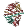
| ||||||||
|---|---|---|---|---|---|---|---|---|---|
| 1 | 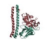
| ||||||||
| 2 | 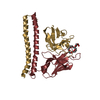
| ||||||||
| Unit cell |
|
-Components
| #1: Antibody | Mass: 20273.049 Da / Num. of mol.: 2 Source method: isolated from a genetically manipulated source Details: MSKIKGHHHHHHGG is derived from plasmid. / Source: (gene. exp.)  #2: Antibody | Mass: 20927.516 Da / Num. of mol.: 2 Source method: isolated from a genetically manipulated source Details: MSKIKGHHHHHHGG is derived from plasmid. / Source: (gene. exp.) #3: Chemical | Mass: 412.516 Da / Num. of mol.: 2 / Source method: isolated from a natural source / Formula: C19H40O9 / Feature type: SUBJECT OF INVESTIGATION #4: Water | ChemComp-HOH / |  Mass: 18.015 Da / Num. of mol.: 126 / Source method: isolated from a natural source / Formula: H2O Mass: 18.015 Da / Num. of mol.: 126 / Source method: isolated from a natural source / Formula: H2OHas ligand of interest | Y | |
|---|
-Experimental details
-Experiment
| Experiment | Method: X-RAY DIFFRACTION / Number of used crystals: 1 |
|---|
- Sample preparation
| Crystal | Density Matthews: 2.42 Å3/Da / Density % sol: 44.11 % |
|---|---|
| Crystal grow | Temperature: 293 K / Method: vapor diffusion, hanging drop / Details: 0.2 M Sodium formate and 16% (v/v) PEG 550DME |
-Data collection
| Diffraction | Mean temperature: 100 K / Serial crystal experiment: N |
|---|---|
| Diffraction source | Source: SYNCHROTRON / Site: SPring-8 / Beamline: BL45XU / Wavelength: 1 Å |
| Detector | Type: DECTRIS PILATUS3 6M / Detector: PIXEL / Date: Oct 16, 2021 |
| Radiation | Protocol: SINGLE WAVELENGTH / Monochromatic (M) / Laue (L): M / Scattering type: x-ray |
| Radiation wavelength | Wavelength: 1 Å / Relative weight: 1 |
| Reflection | Resolution: 2.35→49.4 Å / Num. obs: 29444 / % possible obs: 98.2 % / Redundancy: 3.18 % / CC1/2: 0.997 / Rrim(I) all: 0.108 / Net I/σ(I): 10.36 |
| Reflection shell | Resolution: 2.35→2.49 Å / Mean I/σ(I) obs: 1.56 / Num. unique obs: 4683 / CC1/2: 0.572 / Rrim(I) all: 1.062 |
- Processing
| Software |
| |||||||||||||||||||||||||||||||||||||||||||||||||||||||||||||||||||||||||||||||||||||||||||||||||||||||||
|---|---|---|---|---|---|---|---|---|---|---|---|---|---|---|---|---|---|---|---|---|---|---|---|---|---|---|---|---|---|---|---|---|---|---|---|---|---|---|---|---|---|---|---|---|---|---|---|---|---|---|---|---|---|---|---|---|---|---|---|---|---|---|---|---|---|---|---|---|---|---|---|---|---|---|---|---|---|---|---|---|---|---|---|---|---|---|---|---|---|---|---|---|---|---|---|---|---|---|---|---|---|---|---|---|---|---|
| Refinement | Method to determine structure: MOLECULAR REPLACEMENT / Resolution: 2.35→49.4 Å / SU ML: 0.34 / Cross valid method: FREE R-VALUE / σ(F): 1.35 / Phase error: 33.03 / Stereochemistry target values: ML
| |||||||||||||||||||||||||||||||||||||||||||||||||||||||||||||||||||||||||||||||||||||||||||||||||||||||||
| Solvent computation | Shrinkage radii: 0.9 Å / VDW probe radii: 1.1 Å / Solvent model: FLAT BULK SOLVENT MODEL | |||||||||||||||||||||||||||||||||||||||||||||||||||||||||||||||||||||||||||||||||||||||||||||||||||||||||
| Refinement step | Cycle: LAST / Resolution: 2.35→49.4 Å
| |||||||||||||||||||||||||||||||||||||||||||||||||||||||||||||||||||||||||||||||||||||||||||||||||||||||||
| Refine LS restraints |
| |||||||||||||||||||||||||||||||||||||||||||||||||||||||||||||||||||||||||||||||||||||||||||||||||||||||||
| LS refinement shell |
|
