| 登録情報 | データベース: PDB / ID: 8iqs
|
|---|
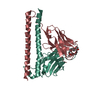
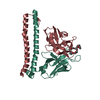
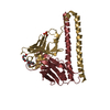
| タイトル | Crystal structure of Anti-PEG antibody M11 Fv-clasp fragment with PEG (co-crystallization with PEG3350) |
|---|
 要素 要素 | |
|---|
 キーワード キーワード | IMMUNE SYSTEM |
|---|
| 機能・相同性 | 3,6,9,12,15,18,21-HEPTAOXATRICOSANE-1,23-DIOL 機能・相同性情報 機能・相同性情報 |
|---|
| 生物種 |   Mus musculus (ハツカネズミ) Mus musculus (ハツカネズミ) |
|---|
| 手法 |  X線回折 / シンクロトロン / 分子置換 / 解像度: 2.16 Å X線回折 / シンクロトロン / 分子置換 / 解像度: 2.16 Å |
|---|
 データ登録者 データ登録者 | Mori, T. / Teramoto, T. / Liu, Y. / Mori, T. / Kakuta, Y. |
|---|
| 資金援助 |  日本, 1件 日本, 1件 | 組織 | 認可番号 | 国 |
|---|
| Japan Society for the Promotion of Science (JSPS) | | 日本 |
|
|---|
 引用 引用 | ジャーナル: J Control Release / 年: 2025
タイトル: The strategy used by naive anti-PEG antibodies to capture flexible and featureless PEG chains.
著者: Liu, Y. / Mori, T. / Ito, Y. / Kuroki, K. / Hayashi, S. / Kohda, D. / Shimizu, T. / Ishida, T. / Roffler, S.R. / Kaneko, M.K. / Kato, Y. / Arimori, T. / Teramoto, T. / Takemura, K. / ...著者: Liu, Y. / Mori, T. / Ito, Y. / Kuroki, K. / Hayashi, S. / Kohda, D. / Shimizu, T. / Ishida, T. / Roffler, S.R. / Kaneko, M.K. / Kato, Y. / Arimori, T. / Teramoto, T. / Takemura, K. / Ishibashi, K. / Katayama, Y. / Maenaka, K. / Kakuta, Y. / Kitao, A. / Mori, T. |
|---|
| 履歴 | | 登録 | 2023年3月17日 | 登録サイト: PDBJ / 処理サイト: PDBJ |
|---|
| 改定 1.0 | 2024年3月20日 | Provider: repository / タイプ: Initial release |
|---|
| 改定 1.1 | 2024年10月9日 | Group: Structure summary
カテゴリ: pdbx_entry_details / pdbx_modification_feature
Item: _pdbx_entry_details.has_protein_modification |
|---|
| 改定 1.2 | 2025年3月19日 | Group: Database references / カテゴリ: citation / citation_author
Item: _citation.journal_abbrev / _citation.journal_id_CSD ..._citation.journal_abbrev / _citation.journal_id_CSD / _citation.journal_id_ISSN / _citation.journal_volume / _citation.page_first / _citation.page_last / _citation.pdbx_database_id_DOI / _citation.title / _citation.year |
|---|
| 改定 1.3 | 2025年3月26日 | Group: Database references / カテゴリ: citation / Item: _citation.pdbx_database_id_PubMed / _citation.title |
|---|
|
|---|
 ムービー
ムービー コントローラー


 データを開く
 基本情報
基本情報 構造の表示
構造の表示 ダウンロードとリンク
 その他のダウンロード
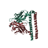
 PDBj
PDBj

 集合体
集合体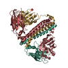


 分子量: 96.063 Da / 分子数: 2 / 由来タイプ: 合成 / 式: SO4 / タイプ: SUBJECT OF INVESTIGATION
 分子量: 370.436 Da / 分子数: 2 / 由来タイプ: 合成 / 式: C16H34O9 / タイプ: SUBJECT OF INVESTIGATION
 分子量: 18.015 Da / 分子数: 86 / 由来タイプ: 天然 / 式: H2O
 試料調製
試料調製 解析
解析