[English] 日本語
Yorodumi
- PDB-8ilu: Crystal structure of mouse Galectin-3 in complex with small molec... -

+
Open data


ID or keywords:

Loading...

-
Basic information

Entry
Database: PDB / ID: 8ilu
TitleCrystal structure of mouse Galectin-3 in complex with small molecule inhibitor
ComponentsGalectin-3
KeywordsSUGAR BINDING PROTEIN / Fibrosis / Galactose
Function / homology
Function and homology information


negative regulation of cell proliferation in bone marrow / advanced glycation end-product receptor activity / positive regulation of serotonin secretion / negative regulation of immunological synapse formation / disaccharide binding / negative regulation of T cell activation via T cell receptor contact with antigen bound to MHC molecule on antigen presenting cell / cornified envelope / IgE binding / negative regulation of endocytosis / Fc-gamma receptor I complex binding ...negative regulation of cell proliferation in bone marrow / advanced glycation end-product receptor activity / positive regulation of serotonin secretion / negative regulation of immunological synapse formation / disaccharide binding / negative regulation of T cell activation via T cell receptor contact with antigen bound to MHC molecule on antigen presenting cell / cornified envelope / IgE binding / negative regulation of endocytosis / Fc-gamma receptor I complex binding / eosinophil chemotaxis / monosaccharide binding / negative regulation of T cell receptor signaling pathway / positive chemotaxis / positive regulation of calcium ion import / macrophage chemotaxis / monocyte chemotaxis / glial cell projection / immunological synapse / laminin binding / Neutrophil degranulation / RNA splicing / neutrophil chemotaxis / extracellular matrix organization / extracellular matrix / skeletal system development / negative regulation of extrinsic apoptotic signaling pathway / spliceosomal complex / mRNA processing / positive regulation of angiogenesis / collagen-containing extracellular matrix / cell differentiation / external side of plasma membrane / innate immune response / positive regulation of cell population proliferation / negative regulation of apoptotic process / cell surface / extracellular space / extracellular region / nucleus / cytoplasm
Similarity search - Function
Galectin-like / Galactoside-binding lectin / Galectin / Galectin, carbohydrate recognition domain / Galactoside-binding lectin / Galactoside-binding lectin (galectin) domain profile. / Concanavalin A-like lectin/glucanase domain superfamily
Similarity search - Domain/homology
: / THIOCYANATE ION / Galectin-3
Similarity search - Component
Biological speciesMus musculus (house mouse)
MethodX-RAY DIFFRACTION / SYNCHROTRON / MOLECULAR REPLACEMENT / Resolution: 1.8 Å
AuthorsKumar, A. / Jinal, S. / Raman, S. / Ghosh, K.
Funding support1items
OrganizationGrant numberCountry
Other private
CitationJournal: To Be Published
Title: Identification of Benzothiazole Derived Monosaccharides as Potent, Selective, and Orally Bioavailable Inhibitors of Human and Mouse Galectin-3
Authors: Ghosh, K.
History
DepositionMar 4, 2023Deposition site: PDBJ / Processing site: PDBJ
Revision 1.0Mar 6, 2024Provider: repository / Type: Initial release

-
Structure visualization

Structure viewerMolecule:
MolmilJmol/JSmol

Downloads & links

-
Assembly

Deposited unit
A: Galectin-3
B: Galectin-3
hetero molecules


Theoretical massNumber of molelcules
Total (without water)34,1106
Polymers32,9102
Non-polymers1,2004
Water3,477193
1


  • Idetical with deposited unit
  • defined by author
  • Evidence: light scattering
TypeNameSymmetry operationNumber
identity operation1_555x,y,z1
Buried area1220 Å2
ΔGint-5 kcal/mol
Surface area12950 Å2
Unit cell
Length a, b, c (Å)40.730, 49.600, 60.550
Angle α, β, γ (deg.)90.00, 94.16, 90.00
Int Tables number4
Space group name H-MP1211

-
Components

#1: Protein Galectin-3 / Gal-3 / 35 kDa lectin / Carbohydrate-binding protein 35 / CBP 35 / Galactose-specific lectin 3 / ...Gal-3 / 35 kDa lectin / Carbohydrate-binding protein 35 / CBP 35 / Galactose-specific lectin 3 / IgE-binding protein / L-34 galactoside-binding lectin / Laminin-binding protein / Lectin L-29 / Mac-2 antigen


Mass: 16454.906 Da / Num. of mol.: 2
Source method: isolated from a genetically manipulated source
Source: (gene. exp.) Mus musculus (house mouse) / Gene: Lgals3 / Production host: Escherichia coli (E. coli) / References: UniProt: P16110
#2: Chemical ChemComp-Q1L / (2R,3R,4R,5R,6S)-2-(hydroxymethyl)-6-[2-(2-methyl-1,3-benzothiazol-6-yl)-1,2,4-triazol-3-yl]-4-[4-[3,4,5-tris(fluoranyl)phenyl]-1,2,3-triazol-1-yl]oxane-3,5-diol


Mass: 559.520 Da / Num. of mol.: 2 / Source method: obtained synthetically / Formula: C24H20F3N7O4S / Feature type: SUBJECT OF INVESTIGATION
#3: Chemical ChemComp-SCN / THIOCYANATE ION


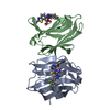
Mass: 58.082 Da / Num. of mol.: 1 / Source method: obtained synthetically / Formula: CNS
#4: Chemical ChemComp-NA / SODIUM ION


Mass: 22.990 Da / Num. of mol.: 1 / Source method: obtained synthetically / Formula: Na
#5: Water ChemComp-HOH / water


Mass: 18.015 Da / Num. of mol.: 193 / Source method: isolated from a natural source / Formula: H2O
Has ligand of interestY

-
Experimental details

-
Experiment

ExperimentMethod: X-RAY DIFFRACTION / Number of used crystals: 1

-
Sample preparation

CrystalDensity Matthews: 1.85 Å3/Da / Density % sol: 33.64 %
Crystal growTemperature: 293 K / Method: vapor diffusion, hanging drop / pH: 8.2 / Details: 30 % PEG 6K, 0.100M TRIS PH8.2, 0.400M NASCN

-
Data collection

DiffractionMean temperature: 100 K / Serial crystal experiment: N
Diffraction sourceSource: SYNCHROTRON / Site: APS / Beamline: 22-ID / Wavelength: 0.969 Å
DetectorType: RAYONIX MX300-HS / Detector: CCD / Date: Jul 30, 2017
RadiationProtocol: SINGLE WAVELENGTH / Monochromatic (M) / Laue (L): M / Scattering type: x-ray
Radiation wavelengthWavelength: 0.969 Å / Relative weight: 1
ReflectionResolution: 1.8→31.43 Å / Num. obs: 21379 / % possible obs: 99.7 % / Redundancy: 3.7 % / Rmerge(I) obs: 0.073 / Net I/σ(I): 10.9
Reflection shellResolution: 1.8→1.847 Å / Num. unique obs: 1602 / Rrim(I) all: 0.206 / % possible all: 100

-
Processing

Software
NameVersionClassification
REFMAC5.8.0049refinement
XDSdata reduction
Aimlessdata scaling
PHASERphasing
RefinementMethod to determine structure: MOLECULAR REPLACEMENT / Resolution: 1.8→31.43 Å / Cor.coef. Fo:Fc: 0.964 / Cor.coef. Fo:Fc free: 0.931 / SU B: 2.956 / SU ML: 0.094 / Cross valid method: THROUGHOUT / ESU R: 0.138 / ESU R Free: 0.142 / Stereochemistry target values: MAXIMUM LIKELIHOOD / Details: HYDROGENS HAVE BEEN USED IF PRESENT IN TH INPUT
RfactorNum. reflection% reflectionSelection details
Rfree0.229 1072 4.8 %RANDOM
Rwork0.164 ---
obs0.167 21379 99.7 %-
Solvent computationIon probe radii: 0.8 Å / Shrinkage radii: 0.8 Å / VDW probe radii: 1.2 Å / Solvent model: MASK
Displacement parametersBiso mean: 21.94 Å2
Baniso -1Baniso -2Baniso -3
1-0.29 Å20 Å20.22 Å2
2--0.53 Å20 Å2
3----0.84 Å2
Refinement stepCycle: LAST / Resolution: 1.8→31.43 Å
ProteinNucleic acidLigandSolventTotal
Num. atoms2166 0 82 193 2441
Refine LS restraints
Refine-IDTypeDev idealNumber
X-RAY DIFFRACTIONr_bond_refined_d0.022327
X-RAY DIFFRACTIONr_bond_other_d040
X-RAY DIFFRACTIONr_angle_refined_deg2.1783174
X-RAY DIFFRACTIONr_angle_other_deg1.01486
X-RAY DIFFRACTIONr_dihedral_angle_1_deg6.822281
X-RAY DIFFRACTIONr_dihedral_angle_2_deg37.312108
X-RAY DIFFRACTIONr_dihedral_angle_3_deg15.717374
X-RAY DIFFRACTIONr_dihedral_angle_4_deg19.1818
X-RAY DIFFRACTIONr_chiral_restr0.165350
X-RAY DIFFRACTIONr_gen_planes_refined0.0122080
X-RAY DIFFRACTIONr_gen_planes_other0.00114
X-RAY DIFFRACTIONr_nbd_refined
X-RAY DIFFRACTIONr_nbd_other
X-RAY DIFFRACTIONr_nbtor_refined
X-RAY DIFFRACTIONr_nbtor_other
X-RAY DIFFRACTIONr_xyhbond_nbd_refined
X-RAY DIFFRACTIONr_xyhbond_nbd_other
X-RAY DIFFRACTIONr_metal_ion_refined
X-RAY DIFFRACTIONr_metal_ion_other
X-RAY DIFFRACTIONr_symmetry_vdw_refined
X-RAY DIFFRACTIONr_symmetry_vdw_other
X-RAY DIFFRACTIONr_symmetry_hbond_refined
X-RAY DIFFRACTIONr_symmetry_hbond_other
X-RAY DIFFRACTIONr_symmetry_metal_ion_refined
X-RAY DIFFRACTIONr_symmetry_metal_ion_other
X-RAY DIFFRACTIONr_mcbond_it2.3161109
X-RAY DIFFRACTIONr_mcbond_other2.3131108
X-RAY DIFFRACTIONr_mcangle_it3.3551383
X-RAY DIFFRACTIONr_mcangle_other
X-RAY DIFFRACTIONr_scbond_it3.9671218
X-RAY DIFFRACTIONr_scbond_other
X-RAY DIFFRACTIONr_scangle_it
X-RAY DIFFRACTIONr_scangle_other
X-RAY DIFFRACTIONr_long_range_B_refined
X-RAY DIFFRACTIONr_long_range_B_other
X-RAY DIFFRACTIONr_rigid_bond_restr
X-RAY DIFFRACTIONr_sphericity_free
X-RAY DIFFRACTIONr_sphericity_bonded
LS refinement shellResolution: 1.8→1.85 Å / Total num. of bins used: 20
RfactorNum. reflection% reflection
Rfree0.264 75 -
Rwork0.206 1602 -
obs--100 %

+
About Yorodumi

-
News

-
Feb 9, 2022. New format data for meta-information of EMDB entries

New format data for meta-information of EMDB entries

  • Version 3 of the EMDB header file is now the official format.
  • The previous official version 1.9 will be removed from the archive.

Related info.:EMDB header

External links:wwPDB to switch to version 3 of the EMDB data model

-
Aug 12, 2020. Covid-19 info

Covid-19 info

URL: https://pdbjlvh1.pdbj.org/emnavi/covid19.php

New page: Covid-19 featured information page in EM Navigator.

Related info.:Covid-19 info / Mar 5, 2020. Novel coronavirus structure data

+
Mar 5, 2020. Novel coronavirus structure data

Novel coronavirus structure data

Related info.:Yorodumi Speices / Aug 12, 2020. Covid-19 info

External links:COVID-19 featured content - PDBj / Molecule of the Month (242):Coronavirus Proteases

+
Jan 31, 2019. EMDB accession codes are about to change! (news from PDBe EMDB page)

EMDB accession codes are about to change! (news from PDBe EMDB page)

  • The allocation of 4 digits for EMDB accession codes will soon come to an end. Whilst these codes will remain in use, new EMDB accession codes will include an additional digit and will expand incrementally as the available range of codes is exhausted. The current 4-digit format prefixed with “EMD-” (i.e. EMD-XXXX) will advance to a 5-digit format (i.e. EMD-XXXXX), and so on. It is currently estimated that the 4-digit codes will be depleted around Spring 2019, at which point the 5-digit format will come into force.
  • The EM Navigator/Yorodumi systems omit the EMD- prefix.

Related info.:Q: What is EMD? / ID/Accession-code notation in Yorodumi/EM Navigator

External links:EMDB Accession Codes are Changing Soon! / Contact to PDBj

+
Jul 12, 2017. Major update of PDB

Major update of PDB

  • wwPDB released updated PDB data conforming to the new PDBx/mmCIF dictionary.
  • This is a major update changing the version number from 4 to 5, and with Remediation, in which all the entries are updated.
  • In this update, many items about electron microscopy experimental information are reorganized (e.g. em_software).
  • Now, EM Navigator and Yorodumi are based on the updated data.

External links:wwPDB Remediation / Enriched Model Files Conforming to OneDep Data Standards Now Available in the PDB FTP Archive

-
Yorodumi

Thousand views of thousand structures

  • Yorodumi is a browser for structure data from EMDB, PDB, SASBDB, etc.
  • This page is also the successor to EM Navigator detail page, and also detail information page/front-end page for Omokage search.
  • The word "yorodu" (or yorozu) is an old Japanese word meaning "ten thousand". "mi" (miru) is to see.

Related info.:EMDB / PDB / SASBDB / Comparison of 3 databanks / Yorodumi Search / Aug 31, 2016. New EM Navigator & Yorodumi / Yorodumi Papers / Jmol/JSmol / Function and homology information / Changes in new EM Navigator and Yorodumi

Read more