 Movie
Movie Controller


+ Open data

- Basic information
| Entry | Database: PDB / ID: 8iir | |||||||||
|---|---|---|---|---|---|---|---|---|---|---|
| Title | MsmUdgX H109S/Q53A double mutant | |||||||||
 Components Components | Type-4 uracil-DNA glycosylase | |||||||||
 Keywords Keywords | DNA BINDING PROTEIN / UdgX / H109S / Q53A / mutant / protein | |||||||||
| Function / homology |  Function and homology information Function and homology informationuracil DNA N-glycosylase activity / 4 iron, 4 sulfur cluster binding / DNA repair / metal ion binding Similarity search - Function | |||||||||
| Biological species |  Mycolicibacterium smegmatis MC2 155 (bacteria) Mycolicibacterium smegmatis MC2 155 (bacteria) | |||||||||
| Method |  X-RAY DIFFRACTION / MOLECULAR REPLACEMENT / Resolution: 2.28 Å X-RAY DIFFRACTION / MOLECULAR REPLACEMENT / Resolution: 2.28 Å | |||||||||
 Authors Authors | Aroli, S. | |||||||||
| Funding support |  India, 2items India, 2items
| |||||||||
 Citation Citation | Journal: Nucleic Acids Res. / Year: 2023 Title: Mutational and structural analyses of UdgX: insights into the active site pocket architecture and its evolution. Authors: Aroli, S. / Woo, E.J. / Gopal, B. / Varshney, U. | |||||||||
| History |
|
- Structure visualization
| Structure viewer | Molecule: MolmilJmol/JSmol |
|---|
- Downloads & links
-Download
| PDBx/mmCIF format | 8iir.cif.gz | 54.6 KB | Display | PDBx/mmCIF format |
|---|---|---|---|---|
| PDB format | pdb8iir.ent.gz | 36.8 KB | Display | PDB format |
| PDBx/mmJSON format | 8iir.json.gz | Tree view | PDBx/mmJSON format | |
| Others |  Other downloads Other downloads |
-Validation report
| Summary document | 8iir_validation.pdf.gz | 1.1 MB | Display | wwPDB validaton report |
|---|---|---|---|---|
| Full document | 8iir_full_validation.pdf.gz | 1.1 MB | Display | |
| Data in XML | 8iir_validation.xml.gz | 10.1 KB | Display | |
| Data in CIF | 8iir_validation.cif.gz | 13.4 KB | Display | |
| Arichive directory | https://data.pdbj.org/pub/pdb/validation_reports/ii/8iirftp://data.pdbj.org/pub/pdb/validation_reports/ii/8iir | HTTPS FTP |
-Related structure data
| Related structure data | 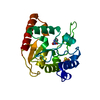 8iieC 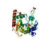 8iifC 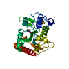 8iigC 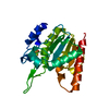 8iihC 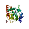 8iiiC 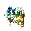 8iijC 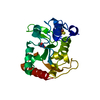 8iilC 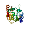 8iimC 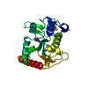 8iinC 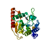 8iioC 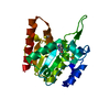 8iipC 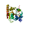 8iiqC 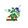 8iisC 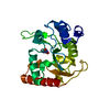 8iitC C: citing same article ( |
|---|---|
| Similar structure data |
-Links
 PDBj
PDBj


- Assembly
| Deposited unit | 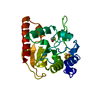
| ||||||||
|---|---|---|---|---|---|---|---|---|---|
| 1 |
| ||||||||
| Unit cell |
|
-Components
| #1: Protein | Mass: 21707.656 Da / Num. of mol.: 1 / Mutation: Q53A,H109S Source method: isolated from a genetically manipulated source Source: (gene. exp.) Mycolicibacterium smegmatis MC2 155 (bacteria)Strain: MC2 155 / Gene: MSMEG_0265 / Plasmid: pET14b / Production host: |
|---|---|
| #2: Chemical | ChemComp-SF4 /   Mass: 351.640 Da / Num. of mol.: 1 / Source method: obtained synthetically / Formula: Fe4S4 / Feature type: SUBJECT OF INVESTIGATION Mass: 351.640 Da / Num. of mol.: 1 / Source method: obtained synthetically / Formula: Fe4S4 / Feature type: SUBJECT OF INVESTIGATION |
| #3: Chemical | ChemComp-BME /   Mass: 78.133 Da / Num. of mol.: 1 / Source method: obtained synthetically / Formula: C2H6OS / Feature type: SUBJECT OF INVESTIGATION Mass: 78.133 Da / Num. of mol.: 1 / Source method: obtained synthetically / Formula: C2H6OS / Feature type: SUBJECT OF INVESTIGATION |
| #4: Water | ChemComp-HOH /  Mass: 18.015 Da / Num. of mol.: 79 / Source method: isolated from a natural source / Formula: H2O Mass: 18.015 Da / Num. of mol.: 79 / Source method: isolated from a natural source / Formula: H2O |
| Has ligand of interest | Y |
-Experimental details
-Experiment
| Experiment | Method: X-RAY DIFFRACTION / Number of used crystals: 1 |
|---|
- Sample preparation
| Crystal | Density Matthews: 2.1 Å3/Da / Density % sol: 46.26 % |
|---|---|
| Crystal grow | Temperature: 295 K / Method: microbatch / pH: 7 Details: 2.0M Ammonium citrate tribasic pH7.0, 0.1M BIS-TRIS propane pH7.0 PH range: 6.8-7.2 |
-Data collection
| Diffraction | Mean temperature: 100 K / Serial crystal experiment: N |
|---|---|
| Diffraction source | Source: ROTATING ANODE / Type: BRUKER AXS MICROSTAR / Wavelength: 1.54179 Å |
| Detector | Type: MAR scanner 345 mm plate / Detector: IMAGE PLATE / Date: Mar 22, 2021 |
| Radiation | Monochromator: M / Protocol: SINGLE WAVELENGTH / Monochromatic (M) / Laue (L): M / Scattering type: x-ray |
| Radiation wavelength | Wavelength: 1.54179 Å / Relative weight: 1 |
| Reflection | Resolution: 2.28→53.23 Å / Num. obs: 9050 / % possible obs: 99.9 % / Redundancy: 3 % / CC1/2: 0.986 / Rmerge(I) obs: 0.098 / Rpim(I) all: 0.096 / Rrim(I) all: 0.137 / Net I/σ(I): 6.2 |
| Reflection shell | Resolution: 2.28→2.37 Å / Redundancy: 3 % / Rmerge(I) obs: 0.315 / Mean I/σ(I) obs: 2.2 / Num. unique obs: 925 / CC1/2: 0.822 / Rpim(I) all: 0.3 / Rrim(I) all: 0.436 / % possible all: 99.9 |
- Processing
| Software |
| ||||||||||||||||||||||||||||
|---|---|---|---|---|---|---|---|---|---|---|---|---|---|---|---|---|---|---|---|---|---|---|---|---|---|---|---|---|---|
| Refinement | Method to determine structure: MOLECULAR REPLACEMENT / Resolution: 2.28→53.23 Å / SU ML: 0.23 / Cross valid method: THROUGHOUT / σ(F): 1.98 / Phase error: 21.75 / Stereochemistry target values: MLHL
| ||||||||||||||||||||||||||||
| Solvent computation | Shrinkage radii: 0.9 Å / VDW probe radii: 1.11 Å / Solvent model: FLAT BULK SOLVENT MODEL | ||||||||||||||||||||||||||||
| Refinement step | Cycle: LAST / Resolution: 2.28→53.23 Å
| ||||||||||||||||||||||||||||
| Refine LS restraints |
| ||||||||||||||||||||||||||||
| LS refinement shell |
|
