| Entry | Database: PDB / ID: 8ifo
|
|---|
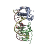
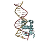
| Title | Crystal structure of estrogen related receptor-gamma DNA binding domain complexed with Pla2g12b promoter |
|---|
 Components Components | - DNA (5'-D(*GP*AP*GP*GP*AP*CP*AP*AP*AP*GP*GP*TP*GP*AP*AP*AP*C)-3')
- DNA (5'-D(*GP*TP*TP*TP*CP*AP*CP*CP*TP*TP*TP*GP*TP*CP*CP*TP*C)-3')
- Estrogen-related receptor gamma
|
|---|
 Keywords Keywords | TRANSCRIPTION / nuclear receptor / ERR3 / PLA2G12B / DR1 |
|---|
| Function / homology |  Function and homology information Function and homology information
AF-2 domain binding / nuclear steroid receptor activity / estrogen response element binding / retinoic acid receptor signaling pathway / steroid binding / Nuclear Receptor transcription pathway / nuclear receptor activity / sequence-specific double-stranded DNA binding / positive regulation of cold-induced thermogenesis / DNA-binding transcription activator activity, RNA polymerase II-specific ...AF-2 domain binding / nuclear steroid receptor activity / estrogen response element binding / retinoic acid receptor signaling pathway / steroid binding / Nuclear Receptor transcription pathway / nuclear receptor activity / sequence-specific double-stranded DNA binding / positive regulation of cold-induced thermogenesis / DNA-binding transcription activator activity, RNA polymerase II-specific / DNA-binding transcription factor activity, RNA polymerase II-specific / regulation of transcription by RNA polymerase II / regulation of DNA-templated transcription / chromatin / positive regulation of transcription by RNA polymerase II / zinc ion binding / nucleoplasm / identical protein binding / nucleusSimilarity search - Function Oestrogen-related receptor / Retinoic acid receptor / Estrogen receptor/oestrogen-related receptor / : / Nuclear hormone receptor / Nuclear hormones receptors DNA-binding region signature. / Zinc finger, nuclear hormone receptor-type / Double treble clef zinc finger, C4 type / Nuclear hormone receptors DNA-binding domain profile. / c4 zinc finger in nuclear hormone receptors ...Oestrogen-related receptor / Retinoic acid receptor / Estrogen receptor/oestrogen-related receptor / : / Nuclear hormone receptor / Nuclear hormones receptors DNA-binding region signature. / Zinc finger, nuclear hormone receptor-type / Double treble clef zinc finger, C4 type / Nuclear hormone receptors DNA-binding domain profile. / c4 zinc finger in nuclear hormone receptors / Nuclear hormone receptor, ligand-binding domain / Nuclear hormone receptor-like domain superfamily / Ligand-binding domain of nuclear hormone receptor / Nuclear receptor (NR) ligand-binding (LBD) domain profile. / Ligand binding domain of hormone receptors / Zinc finger, NHR/GATA-typeSimilarity search - Domain/homology |
|---|
| Biological species |  Homo sapiens (human) Homo sapiens (human)
  Mus musculus (house mouse) Mus musculus (house mouse) |
|---|
| Method |  X-RAY DIFFRACTION / SYNCHROTRON / MOLECULAR REPLACEMENT / Resolution: 2.2 Å X-RAY DIFFRACTION / SYNCHROTRON / MOLECULAR REPLACEMENT / Resolution: 2.2 Å |
|---|
 Authors Authors | Xu, T. / Zhen, X. / Liu, J. |
|---|
| Funding support | 1items | Organization | Grant number | Country |
|---|
| Not funded | | |
|
|---|
 Citation Citation | Journal: Biochem.Biophys.Res.Commun. / Year: 2023
Title: ERR gamma-DBD undergoes dimerization and conformational rearrangement upon binding to the downstream site of the DR1 element.
Authors: Zhen, X. / Gan, Q. / Qu, L. / Dong, Y. / Pan, C. / Liu, J. / Wang, N. / Xu, T. |
|---|
| History | | Deposition | Feb 19, 2023 | Deposition site: PDBJ / Processing site: PDBC |
|---|
| Revision 1.0 | Mar 29, 2023 | Provider: repository / Type: Initial release |
|---|
| Revision 1.1 | May 1, 2024 | Group: Data collection / Database references / Category: chem_comp_atom / chem_comp_bond / citation / Item: _citation.pdbx_database_id_PubMed / _citation.title |
|---|
|
|---|
 Movie
Movie Controller

 Yorodumi
 Open data
 Basic information
 Structure visualization
 Downloads & links
 Other downloads
 PDBj
PDBj


 Assembly
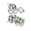


 Mass: 65.409 Da / Num. of mol.: 7 / Source method: isolated from a natural source / Formula: Zn / Feature type: SUBJECT OF INVESTIGATION
 Mass: 102.046 Da / Num. of mol.: 3 / Source method: obtained synthetically / Formula: C3H2O4
 Sample preparation
 / Beamline: BL19U1 / Wavelength: 0.9785 Å
 Processing