 Movie
Movie Controller

[English] 日本語
 Yorodumi
- PDB-8i28: Structure of Phosphoserine Aminotransferase from Saccharomyces ce... -
+ Open data

- Basic information
| Entry | Database: PDB / ID: 8i28 | ||||||
|---|---|---|---|---|---|---|---|
| Title | Structure of Phosphoserine Aminotransferase from Saccharomyces cerevisiae | ||||||
 Components Components | Phosphoserine aminotransferase | ||||||
 Keywords Keywords | TRANSFERASE / Phosphoserine | ||||||
| Function / homology |  Function and homology information Function and homology informationSerine metabolism / serine family amino acid biosynthetic process / phosphoserine transaminase / O-phospho-L-serine:2-oxoglutarate aminotransferase activity / purine nucleobase biosynthetic process / L-serine biosynthetic process / pyridoxal phosphate binding / cytoplasm Similarity search - Function | ||||||
| Biological species |  | ||||||
| Method |  X-RAY DIFFRACTION / SYNCHROTRON / MOLECULAR REPLACEMENT / Resolution: 2.8 Å X-RAY DIFFRACTION / SYNCHROTRON / MOLECULAR REPLACEMENT / Resolution: 2.8 Å | ||||||
 Authors Authors | Jang, J.Y. / Chang, J.H. | ||||||
| Funding support |  Korea, Republic Of, 1items Korea, Republic Of, 1items
| ||||||
 Citation Citation | Journal: Int J Mol Sci / Year: 2023 Title: Molecular Structure of Phosphoserine Aminotransferase from Saccharomyces cerevisiae. Authors: Jang, J. / Chang, J.H. | ||||||
| History |
|
- Structure visualization
| Structure viewer | Molecule: MolmilJmol/JSmol |
|---|
- Downloads & links
-Download
| PDBx/mmCIF format | 8i28.cif.gz | 270.1 KB | Display | PDBx/mmCIF format |
|---|---|---|---|---|
| PDB format | pdb8i28.ent.gz | 220 KB | Display | PDB format |
| PDBx/mmJSON format | 8i28.json.gz | Tree view | PDBx/mmJSON format | |
| Others |  Other downloads Other downloads |
-Validation report
| Summary document | 8i28_validation.pdf.gz | 443 KB | Display | wwPDB validaton report |
|---|---|---|---|---|
| Full document | 8i28_full_validation.pdf.gz | 457.2 KB | Display | |
| Data in XML | 8i28_validation.xml.gz | 25.8 KB | Display | |
| Data in CIF | 8i28_validation.cif.gz | 35.2 KB | Display | |
| Arichive directory | https://data.pdbj.org/pub/pdb/validation_reports/i2/8i28ftp://data.pdbj.org/pub/pdb/validation_reports/i2/8i28 | HTTPS FTP |
-Related structure data
| Related structure data | 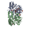 6czxS S: Starting model for refinement |
|---|---|
| Similar structure data |
-Links
 PDBj
PDBj- Assembly
| Deposited unit | 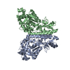
| ||||||||
|---|---|---|---|---|---|---|---|---|---|
| 1 | 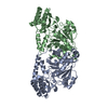
| ||||||||
| Unit cell |
|
-Components
| #1: Protein | Mass: 43470.430 Da / Num. of mol.: 2 Source method: isolated from a genetically manipulated source Source: (gene. exp.) Gene: SER1, SERC, YOR184W / Production host:  #2: Water | ChemComp-HOH / |  Mass: 18.015 Da / Num. of mol.: 53 / Source method: isolated from a natural source / Formula: H2O Mass: 18.015 Da / Num. of mol.: 53 / Source method: isolated from a natural source / Formula: H2OHas ligand of interest | N | |
|---|
-Experimental details
-Experiment
| Experiment | Method: X-RAY DIFFRACTION / Number of used crystals: 1 |
|---|
- Sample preparation
| Crystal | Density Matthews: 4.08 Å3/Da / Density % sol: 69.83 % |
|---|---|
| Crystal grow | Temperature: 293 K / Method: vapor diffusion, hanging drop Details: 2.4 M ammonium sulfate and 0.1 M citric acid (pH 3.5) |
-Data collection
| Diffraction | Mean temperature: 100 K / Serial crystal experiment: N |
|---|---|
| Diffraction source | Source: SYNCHROTRON / Site: PAL/PLS / Beamline: 5C (4A) / Wavelength: 1 Å |
| Detector | Type: ADSC QUANTUM 315r / Detector: CCD / Date: Aug 3, 2020 |
| Radiation | Protocol: SINGLE WAVELENGTH / Monochromatic (M) / Laue (L): M / Scattering type: x-ray |
| Radiation wavelength | Wavelength: 1 Å / Relative weight: 1 |
| Reflection | Resolution: 2.8→50 Å / Num. obs: 35761 / % possible obs: 100 % / Redundancy: 13.4 % / CC1/2: 0.999 / Net I/σ(I): 18.75 |
| Reflection shell | Resolution: 2.8→2.9 Å / Num. unique obs: 3519 / CC1/2: 0.506 |
- Processing
| Software |
| |||||||||||||||||||||||||||||||||||||||||||||||||||||||||||||||||||||||||||||||||||||||||||||||||||||||||
|---|---|---|---|---|---|---|---|---|---|---|---|---|---|---|---|---|---|---|---|---|---|---|---|---|---|---|---|---|---|---|---|---|---|---|---|---|---|---|---|---|---|---|---|---|---|---|---|---|---|---|---|---|---|---|---|---|---|---|---|---|---|---|---|---|---|---|---|---|---|---|---|---|---|---|---|---|---|---|---|---|---|---|---|---|---|---|---|---|---|---|---|---|---|---|---|---|---|---|---|---|---|---|---|---|---|---|
| Refinement | Method to determine structure: MOLECULAR REPLACEMENT Starting model: 6CZX Resolution: 2.8→48.33 Å / SU ML: 0.44 / Cross valid method: FREE R-VALUE / σ(F): 1.5 / Phase error: 28 / Stereochemistry target values: ML
| |||||||||||||||||||||||||||||||||||||||||||||||||||||||||||||||||||||||||||||||||||||||||||||||||||||||||
| Solvent computation | Shrinkage radii: 0.9 Å / VDW probe radii: 1.11 Å / Solvent model: FLAT BULK SOLVENT MODEL | |||||||||||||||||||||||||||||||||||||||||||||||||||||||||||||||||||||||||||||||||||||||||||||||||||||||||
| Refinement step | Cycle: LAST / Resolution: 2.8→48.33 Å
| |||||||||||||||||||||||||||||||||||||||||||||||||||||||||||||||||||||||||||||||||||||||||||||||||||||||||
| Refine LS restraints |
| |||||||||||||||||||||||||||||||||||||||||||||||||||||||||||||||||||||||||||||||||||||||||||||||||||||||||
| LS refinement shell |
| |||||||||||||||||||||||||||||||||||||||||||||||||||||||||||||||||||||||||||||||||||||||||||||||||||||||||
| Refinement TLS params. | Method: refined / Origin x: 74.7884 Å / Origin y: 34.3092 Å / Origin z: 71.4581 Å
| |||||||||||||||||||||||||||||||||||||||||||||||||||||||||||||||||||||||||||||||||||||||||||||||||||||||||
| Refinement TLS group | Selection details: all |
