 Movie
Movie Controller

[English] 日本語
 Yorodumi
- PDB-8i1n: Crystal structure of APSK2 domain from human PAPSS2 in complex wi... -
+ Open data

- Basic information
| Entry | Database: PDB / ID: 8i1n | ||||||
|---|---|---|---|---|---|---|---|
| Title | Crystal structure of APSK2 domain from human PAPSS2 in complex with endogenous APS and ADP | ||||||
 Components Components | Bifunctional 3'-phosphoadenosine 5'-phosphosulfate synthase 2 | ||||||
 Keywords Keywords | BIOSYNTHETIC PROTEIN / Adenosine 5'-phosphosulfate kinase domain 2 / APSK2 / human PAPSS2 | ||||||
| Function / homology |  Function and homology information Function and homology informationDefective PAPSS2 causes SEMD-PA / 3'-phosphoadenosine 5'-phosphosulfate biosynthetic process / Transport and synthesis of PAPS / adenylyl-sulfate kinase / sulfate adenylyltransferase / adenylylsulfate kinase activity / sulfate adenylyltransferase (ATP) activity / Metabolism of ingested H2SeO4 and H2SeO3 into H2Se / sulfate assimilation / hormone metabolic process ...Defective PAPSS2 causes SEMD-PA / 3'-phosphoadenosine 5'-phosphosulfate biosynthetic process / Transport and synthesis of PAPS / adenylyl-sulfate kinase / sulfate adenylyltransferase / adenylylsulfate kinase activity / sulfate adenylyltransferase (ATP) activity / Metabolism of ingested H2SeO4 and H2SeO3 into H2Se / sulfate assimilation / hormone metabolic process / nucleotidyltransferase activity / bone development / blood coagulation / ATP binding / cytosol Similarity search - Function | ||||||
| Biological species |  Homo sapiens (human) Homo sapiens (human) | ||||||
| Method |  X-RAY DIFFRACTION / SYNCHROTRON / MOLECULAR REPLACEMENT / Resolution: 2.8 Å X-RAY DIFFRACTION / SYNCHROTRON / MOLECULAR REPLACEMENT / Resolution: 2.8 Å | ||||||
 Authors Authors | Zhang, L. / Song, W.Y. / Zhang, L. | ||||||
| Funding support |  China, 1items China, 1items
| ||||||
 Citation Citation | Journal: Structure / Year: 2023 Title: Redox switching mechanism of the adenosine 5'-phosphosulfate kinase domain (APSK2) of human PAPS synthase 2. Authors: Zhang, L. / Song, W. / Li, T. / Mu, Y. / Zhang, P. / Hu, J. / Lin, H. / Zhang, J. / Gao, H. / Zhang, L. | ||||||
| History |
|
- Structure visualization
| Structure viewer | Molecule: MolmilJmol/JSmol |
|---|
- Downloads & links
-Download
| PDBx/mmCIF format | 8i1n.cif.gz | 160.1 KB | Display | PDBx/mmCIF format |
|---|---|---|---|---|
| PDB format | pdb8i1n.ent.gz | 129.7 KB | Display | PDB format |
| PDBx/mmJSON format | 8i1n.json.gz | Tree view | PDBx/mmJSON format | |
| Others |  Other downloads Other downloads |
-Validation report
| Arichive directory | https://data.pdbj.org/pub/pdb/validation_reports/i1/8i1nftp://data.pdbj.org/pub/pdb/validation_reports/i1/8i1n | HTTPS FTP |
|---|
-Related structure data
| Related structure data | 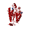 8i1mC  8i1oC  2ofxS S: Starting model for refinement C: citing same article ( |
|---|---|
| Similar structure data |
-Links
 PDBj
PDBj
- Assembly
| Deposited unit | 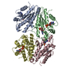
| ||||||||
|---|---|---|---|---|---|---|---|---|---|
| 1 | 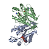
| ||||||||
| 2 | 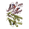
| ||||||||
| Unit cell |
|
-Components
| #1: Protein | Mass: 21809.680 Da / Num. of mol.: 4 / Fragment: APSK2 homodimer domain Source method: isolated from a genetically manipulated source Source: (gene. exp.) Homo sapiens (human) / Gene: PAPSS2, ATPSK2 / Production host:  References: UniProt: O95340, sulfate adenylyltransferase, adenylyl-sulfate kinase #2: Chemical | ChemComp-ADP /   Mass: 427.201 Da / Num. of mol.: 4 / Source method: obtained synthetically / Formula: C10H15N5O10P2 / Comment: ADP, energy-carrying molecule*YM Mass: 427.201 Da / Num. of mol.: 4 / Source method: obtained synthetically / Formula: C10H15N5O10P2 / Comment: ADP, energy-carrying molecule*YM#3: Chemical |   Type: RNA linking / Mass: 427.284 Da / Num. of mol.: 2 / Source method: obtained synthetically / Formula: C10H14N5O10PS / Feature type: SUBJECT OF INVESTIGATION Type: RNA linking / Mass: 427.284 Da / Num. of mol.: 2 / Source method: obtained synthetically / Formula: C10H14N5O10PS / Feature type: SUBJECT OF INVESTIGATIONHas ligand of interest | Y | |
|---|
-Experimental details
-Experiment
| Experiment | Method: X-RAY DIFFRACTION / Number of used crystals: 1 |
|---|
- Sample preparation
| Crystal | Density Matthews: 2.52 Å3/Da / Density % sol: 51.27 % |
|---|---|
| Crystal grow | Temperature: 291 K / Method: vapor diffusion, sitting drop Details: 0.2 M Ammonium citrate dihydrate, pH7.0 and 20% w/v PEG3350 |
-Data collection
| Diffraction | Mean temperature: 100 K / Serial crystal experiment: N | |||||||||||||||||||||||||||||||||||||||||||||||||||||||||||||||||||||||||||||
|---|---|---|---|---|---|---|---|---|---|---|---|---|---|---|---|---|---|---|---|---|---|---|---|---|---|---|---|---|---|---|---|---|---|---|---|---|---|---|---|---|---|---|---|---|---|---|---|---|---|---|---|---|---|---|---|---|---|---|---|---|---|---|---|---|---|---|---|---|---|---|---|---|---|---|---|---|---|---|
| Diffraction source | Source: SYNCHROTRON / Site: SSRF / Beamline: BL19U1 / Wavelength: 0.9875 Å | |||||||||||||||||||||||||||||||||||||||||||||||||||||||||||||||||||||||||||||
| Detector | Type: DECTRIS PILATUS3 6M / Detector: PIXEL / Date: Jan 5, 2021 | |||||||||||||||||||||||||||||||||||||||||||||||||||||||||||||||||||||||||||||
| Radiation | Protocol: SINGLE WAVELENGTH / Monochromatic (M) / Laue (L): M / Scattering type: x-ray | |||||||||||||||||||||||||||||||||||||||||||||||||||||||||||||||||||||||||||||
| Radiation wavelength | Wavelength: 0.9875 Å / Relative weight: 1 | |||||||||||||||||||||||||||||||||||||||||||||||||||||||||||||||||||||||||||||
| Reflection | Resolution: 2.8→50 Å / Num. obs: 21315 / % possible obs: 99 % / Redundancy: 6.2 % / Rmerge(I) obs: 0.974 / Χ2: 0.097 / Net I/σ(I): 3.8 / Num. measured all: 132290 | |||||||||||||||||||||||||||||||||||||||||||||||||||||||||||||||||||||||||||||
| Reflection shell |
|
- Processing
| Software |
| ||||||||||||||||||||||||||||||||||||||||||||||||||||||||
|---|---|---|---|---|---|---|---|---|---|---|---|---|---|---|---|---|---|---|---|---|---|---|---|---|---|---|---|---|---|---|---|---|---|---|---|---|---|---|---|---|---|---|---|---|---|---|---|---|---|---|---|---|---|---|---|---|---|
| Refinement | Method to determine structure: MOLECULAR REPLACEMENT Starting model: 2OFX Resolution: 2.8→44.19 Å / SU ML: 0.34 / Cross valid method: FREE R-VALUE / σ(F): 1.34 / Phase error: 29.16 / Stereochemistry target values: ML
| ||||||||||||||||||||||||||||||||||||||||||||||||||||||||
| Solvent computation | Shrinkage radii: 0.9 Å / VDW probe radii: 1.11 Å / Solvent model: FLAT BULK SOLVENT MODEL | ||||||||||||||||||||||||||||||||||||||||||||||||||||||||
| Refinement step | Cycle: LAST / Resolution: 2.8→44.19 Å
| ||||||||||||||||||||||||||||||||||||||||||||||||||||||||
| Refine LS restraints |
| ||||||||||||||||||||||||||||||||||||||||||||||||||||||||
| LS refinement shell |
|
