 Movie
Movie Controller


+ Open data

- Basic information
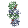
| Entry | Database: PDB / ID: 8hwy | ||||||
|---|---|---|---|---|---|---|---|
| Title | Ancestral imine reductase mutant N559_M6 | ||||||
 Components Components | ancestral imine reductase mutant N559_M6 | ||||||
 Keywords Keywords | OXIDOREDUCTASE / ancestral enzyme / NADP+ | ||||||
| Function / homology | NADP NICOTINAMIDE-ADENINE-DINUCLEOTIDE PHOSPHATE Function and homology information Function and homology information | ||||||
| Biological species |  | ||||||
| Method |  X-RAY DIFFRACTION / SYNCHROTRON / MOLECULAR REPLACEMENT / Resolution: 2.32 Å X-RAY DIFFRACTION / SYNCHROTRON / MOLECULAR REPLACEMENT / Resolution: 2.32 Å | ||||||
 Authors Authors | Zhu, X.X. | ||||||
| Funding support |  China, 1items China, 1items
| ||||||
 Citation Citation | Journal: Nat Commun / Year: 2024 Title: Evolutionary insights into the stereoselectivity of imine reductases based on ancestral sequence reconstruction. Authors: Zhu, X.X. / Zheng, W.Q. / Xia, Z.W. / Chen, X.R. / Jin, T. / Ding, X.W. / Chen, F.F. / Chen, Q. / Xu, J.H. / Kong, X.D. / Zheng, G.W. | ||||||
| History |
|
- Structure visualization
| Structure viewer | Molecule: MolmilJmol/JSmol |
|---|
- Downloads & links
-Download
| PDBx/mmCIF format | 8hwy.cif.gz | 121.9 KB | Display | PDBx/mmCIF format |
|---|---|---|---|---|
| PDB format | pdb8hwy.ent.gz | 93.2 KB | Display | PDB format |
| PDBx/mmJSON format | 8hwy.json.gz | Tree view | PDBx/mmJSON format | |
| Others |  Other downloads Other downloads |
-Validation report
| Arichive directory | https://data.pdbj.org/pub/pdb/validation_reports/hw/8hwyftp://data.pdbj.org/pub/pdb/validation_reports/hw/8hwy | HTTPS FTP |
|---|
-Related structure data
-Links
 PDBj
PDBj- Assembly
| Deposited unit | 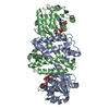
| ||||||||
|---|---|---|---|---|---|---|---|---|---|
| 1 |
| ||||||||
| Unit cell |
|
-Components
| #1: Protein | Mass: 30868.789 Da / Num. of mol.: 2 Source method: isolated from a genetically manipulated source Details: Sequence reference for this entry is not available in UniProt at the time of biocuration. Source: (gene. exp.) #2: Chemical |   Mass: 743.405 Da / Num. of mol.: 2 / Source method: obtained synthetically / Formula: C21H28N7O17P3 / Feature type: SUBJECT OF INVESTIGATION Mass: 743.405 Da / Num. of mol.: 2 / Source method: obtained synthetically / Formula: C21H28N7O17P3 / Feature type: SUBJECT OF INVESTIGATION#3: Water | ChemComp-HOH / |  Mass: 18.015 Da / Num. of mol.: 147 / Source method: isolated from a natural source / Formula: H2O Mass: 18.015 Da / Num. of mol.: 147 / Source method: isolated from a natural source / Formula: H2OHas ligand of interest | Y | Has protein modification | N | |
|---|
-Experimental details
-Experiment
| Experiment | Method: X-RAY DIFFRACTION / Number of used crystals: 1 |
|---|
- Sample preparation
| Crystal | Density Matthews: 2.1 Å3/Da / Density % sol: 41.39 % |
|---|---|
| Crystal grow | Temperature: 277.15 K / Method: vapor diffusion, sitting drop / Details: 25% PEG3350, 0.1M Bis-Tris (pH = 6.5), 0.2 M NaCl |
-Data collection
| Diffraction | Mean temperature: 100 K / Serial crystal experiment: N |
|---|---|
| Diffraction source | Source: SYNCHROTRON / Site: SSRF / Beamline: BL19U1 / Wavelength: 0.97853 Å |
| Detector | Type: DECTRIS PILATUS3 6M / Detector: PIXEL / Date: Nov 28, 2022 |
| Radiation | Monochromator: Si11 / Protocol: SINGLE WAVELENGTH / Monochromatic (M) / Laue (L): M / Scattering type: x-ray |
| Radiation wavelength | Wavelength: 0.97853 Å / Relative weight: 1 |
| Reflection | Resolution: 2.32→59.84 Å / Num. obs: 22088 / % possible obs: 99.3 % / Redundancy: 5.9 % / CC1/2: 0.997 / Net I/σ(I): 15.5 |
| Reflection shell | Resolution: 2.32→2.38 Å / Redundancy: 3.9 % / Num. unique obs: 5668 / CC1/2: 0.957 |
- Processing
| Software |
| |||||||||||||||||||||||||||||||||||||||||||||||||||||||||||||||
|---|---|---|---|---|---|---|---|---|---|---|---|---|---|---|---|---|---|---|---|---|---|---|---|---|---|---|---|---|---|---|---|---|---|---|---|---|---|---|---|---|---|---|---|---|---|---|---|---|---|---|---|---|---|---|---|---|---|---|---|---|---|---|---|---|
| Refinement | Method to determine structure: MOLECULAR REPLACEMENT / Resolution: 2.32→52.2 Å / SU ML: 0.24 / Cross valid method: FREE R-VALUE / σ(F): 1.39 / Phase error: 23.3 / Stereochemistry target values: ML
| |||||||||||||||||||||||||||||||||||||||||||||||||||||||||||||||
| Solvent computation | Shrinkage radii: 0.9 Å / VDW probe radii: 1.1 Å / Solvent model: FLAT BULK SOLVENT MODEL | |||||||||||||||||||||||||||||||||||||||||||||||||||||||||||||||
| Refinement step | Cycle: LAST / Resolution: 2.32→52.2 Å
| |||||||||||||||||||||||||||||||||||||||||||||||||||||||||||||||
| Refine LS restraints |
| |||||||||||||||||||||||||||||||||||||||||||||||||||||||||||||||
| LS refinement shell |
|
