 Movie
Movie Controller


+ Open data

- Basic information
| Entry | Database: PDB / ID: 8hu6 | ||||||
|---|---|---|---|---|---|---|---|
| Title | AMP deaminase 2 in complex with AMP | ||||||
 Components Components | AMP deaminase 2 | ||||||
 Keywords Keywords | HYDROLASE / Complex / Deaminase / AMP / IMP / Energy metabolism | ||||||
| Function / homology |  Function and homology information Function and homology informationcyclic purine nucleotide metabolic process / AMP deaminase / AMP deaminase activity / IMP biosynthetic process / AMP metabolic process / podocyte development / Purine salvage / GMP salvage / IMP salvage / GTP metabolic process ...cyclic purine nucleotide metabolic process / AMP deaminase / AMP deaminase activity / IMP biosynthetic process / AMP metabolic process / podocyte development / Purine salvage / GMP salvage / IMP salvage / GTP metabolic process / ATP metabolic process / energy homeostasis / cholesterol homeostasis / metal ion binding / identical protein binding / cytosol Similarity search - Function | ||||||
| Biological species |  Homo sapiens (human) Homo sapiens (human) | ||||||
| Method |  X-RAY DIFFRACTION / SYNCHROTRON / MOLECULAR REPLACEMENT / Resolution: 2.33 Å X-RAY DIFFRACTION / SYNCHROTRON / MOLECULAR REPLACEMENT / Resolution: 2.33 Å | ||||||
 Authors Authors | Adachi, T. / Doi, S. | ||||||
| Funding support | 1items
| ||||||
 Citation Citation | Journal: Bioorg.Med.Chem.Lett. / Year: 2023 Title: The discovery of 3,3-dimethyl-1,2,3,4-tetrahydroquinoxaline-1-carboxamides as AMPD2 inhibitors with a novel mechanism of action. Authors: Kitao, Y. / Saito, T. / Watanabe, S. / Ohe, Y. / Takahashi, K. / Akaki, T. / Adachi, T. / Doi, S. / Yamanaka, K. / Murai, Y. / Oba, M. / Suzuki, T. | ||||||
| History |
|
- Structure visualization
| Structure viewer | Molecule: MolmilJmol/JSmol |
|---|
- Downloads & links
-Download
| PDBx/mmCIF format | 8hu6.cif.gz | 1.1 MB | Display | PDBx/mmCIF format |
|---|---|---|---|---|
| PDB format | pdb8hu6.ent.gz | 787.9 KB | Display | PDB format |
| PDBx/mmJSON format | 8hu6.json.gz | Tree view | PDBx/mmJSON format | |
| Others |  Other downloads Other downloads |
-Validation report
| Arichive directory | https://data.pdbj.org/pub/pdb/validation_reports/hu/8hu6ftp://data.pdbj.org/pub/pdb/validation_reports/hu/8hu6 | HTTPS FTP |
|---|
-Related structure data
| Related structure data | 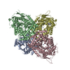 8hubC  2a3lS S: Starting model for refinement C: citing same article ( |
|---|---|
| Similar structure data |
-Links
 PDBj
PDBj


- Assembly
| Deposited unit | 
| ||||||||||||
|---|---|---|---|---|---|---|---|---|---|---|---|---|---|
| 1 |
| ||||||||||||
| Unit cell |
|
-Components
| #1: Protein | Mass: 78752.648 Da / Num. of mol.: 4 Source method: isolated from a genetically manipulated source Source: (gene. exp.) Homo sapiens (human) / Gene: AMPD2 / Production host:   Spodoptera frugiperda (fall armyworm) / References: UniProt: Q01433, AMP deaminase Spodoptera frugiperda (fall armyworm) / References: UniProt: Q01433, AMP deaminase#2: Chemical | ChemComp-ZN /   Mass: 65.409 Da / Num. of mol.: 4 / Source method: obtained synthetically / Formula: Zn Mass: 65.409 Da / Num. of mol.: 4 / Source method: obtained synthetically / Formula: Zn#3: Chemical | ChemComp-SO4 /   Mass: 96.063 Da / Num. of mol.: 4 / Source method: obtained synthetically / Formula: SO4 Mass: 96.063 Da / Num. of mol.: 4 / Source method: obtained synthetically / Formula: SO4#4: Chemical | ChemComp-AMP / |   Mass: 347.221 Da / Num. of mol.: 1 / Source method: obtained synthetically / Formula: C10H14N5O7P / Feature type: SUBJECT OF INVESTIGATION / Comment: AMP*YM Mass: 347.221 Da / Num. of mol.: 1 / Source method: obtained synthetically / Formula: C10H14N5O7P / Feature type: SUBJECT OF INVESTIGATION / Comment: AMP*YM#5: Water | ChemComp-HOH / |  Mass: 18.015 Da / Num. of mol.: 780 / Source method: isolated from a natural source / Formula: H2O Mass: 18.015 Da / Num. of mol.: 780 / Source method: isolated from a natural source / Formula: H2OHas ligand of interest | Y | |
|---|
-Experimental details
-Experiment
| Experiment | Method: X-RAY DIFFRACTION / Number of used crystals: 1 |
|---|
- Sample preparation
| Crystal | Density Matthews: 2.32 Å3/Da / Density % sol: 47.04 % |
|---|---|
| Crystal grow | Temperature: 295.15 K / Method: vapor diffusion, hanging drop / pH: 5.9 Details: 85 mM MES ph 5.9, 20% PEG8000, 170 mM ammonium sulfate, 15% glycerol, 10mM AMP |
-Data collection
| Diffraction | Mean temperature: 100 K / Serial crystal experiment: N |
|---|---|
| Diffraction source | Source: SYNCHROTRON / Site: APS  / Beamline: 22-ID / Wavelength: 1 Å / Beamline: 22-ID / Wavelength: 1 Å |
| Detector | Type: MARMOSAIC 300 mm CCD / Detector: CCD / Date: Jul 27, 2009 |
| Radiation | Protocol: SINGLE WAVELENGTH / Monochromatic (M) / Laue (L): M / Scattering type: x-ray |
| Radiation wavelength | Wavelength: 1 Å / Relative weight: 1 |
| Reflection | Resolution: 2.33→289.39 Å / Num. obs: 122956 / % possible obs: 99 % / Redundancy: 4.1 % / Biso Wilson estimate: 35.54 Å2 / CC1/2: 0.98 / Rmerge(I) obs: 0.08 / Rpim(I) all: 0.049 / Rrim(I) all: 0.094 / Net I/σ(I): 12.1 |
| Reflection shell | Resolution: 2.33→2.39 Å / Rmerge(I) obs: 0.373 / Mean I/σ(I) obs: 3.1 / Num. unique obs: 8943 / CC1/2: 0.91 / Rpim(I) all: 0.189 / Rrim(I) all: 0.42 |
- Processing
| Software |
| |||||||||||||||||||||||||||||||||||||||||||||||||||||||||||||||||||||||||||||||||||||||||||||||||||||||||||||||||||||||||||||||||||||||||||||||||||||||||||||||||||||||||||||||||||||||||||||||||||||||||||||||||||||||||
|---|---|---|---|---|---|---|---|---|---|---|---|---|---|---|---|---|---|---|---|---|---|---|---|---|---|---|---|---|---|---|---|---|---|---|---|---|---|---|---|---|---|---|---|---|---|---|---|---|---|---|---|---|---|---|---|---|---|---|---|---|---|---|---|---|---|---|---|---|---|---|---|---|---|---|---|---|---|---|---|---|---|---|---|---|---|---|---|---|---|---|---|---|---|---|---|---|---|---|---|---|---|---|---|---|---|---|---|---|---|---|---|---|---|---|---|---|---|---|---|---|---|---|---|---|---|---|---|---|---|---|---|---|---|---|---|---|---|---|---|---|---|---|---|---|---|---|---|---|---|---|---|---|---|---|---|---|---|---|---|---|---|---|---|---|---|---|---|---|---|---|---|---|---|---|---|---|---|---|---|---|---|---|---|---|---|---|---|---|---|---|---|---|---|---|---|---|---|---|---|---|---|---|---|---|---|---|---|---|---|---|---|---|---|---|---|---|---|---|
| Refinement | Method to determine structure: MOLECULAR REPLACEMENT Starting model: 2A3L Resolution: 2.33→72.35 Å / SU ML: 0.3407 / Cross valid method: FREE R-VALUE / σ(F): 1.35 / Phase error: 29.6919 Stereochemistry target values: GeoStd + Monomer Library + CDL v1.2
| |||||||||||||||||||||||||||||||||||||||||||||||||||||||||||||||||||||||||||||||||||||||||||||||||||||||||||||||||||||||||||||||||||||||||||||||||||||||||||||||||||||||||||||||||||||||||||||||||||||||||||||||||||||||||
| Solvent computation | Shrinkage radii: 0.9 Å / VDW probe radii: 1.11 Å / Solvent model: FLAT BULK SOLVENT MODEL | |||||||||||||||||||||||||||||||||||||||||||||||||||||||||||||||||||||||||||||||||||||||||||||||||||||||||||||||||||||||||||||||||||||||||||||||||||||||||||||||||||||||||||||||||||||||||||||||||||||||||||||||||||||||||
| Displacement parameters | Biso mean: 43.39 Å2 | |||||||||||||||||||||||||||||||||||||||||||||||||||||||||||||||||||||||||||||||||||||||||||||||||||||||||||||||||||||||||||||||||||||||||||||||||||||||||||||||||||||||||||||||||||||||||||||||||||||||||||||||||||||||||
| Refinement step | Cycle: LAST / Resolution: 2.33→72.35 Å
| |||||||||||||||||||||||||||||||||||||||||||||||||||||||||||||||||||||||||||||||||||||||||||||||||||||||||||||||||||||||||||||||||||||||||||||||||||||||||||||||||||||||||||||||||||||||||||||||||||||||||||||||||||||||||
| Refine LS restraints |
| |||||||||||||||||||||||||||||||||||||||||||||||||||||||||||||||||||||||||||||||||||||||||||||||||||||||||||||||||||||||||||||||||||||||||||||||||||||||||||||||||||||||||||||||||||||||||||||||||||||||||||||||||||||||||
| LS refinement shell |
|
