 Movie
Movie Controller

[English] 日本語
 Yorodumi
- PDB-8hsn: Crystal structure of DFA I-forming Inulin Lyase from Streptomyces... -
+ Open data

- Basic information
| Entry | Database: PDB / ID: 8hsn | ||||||
|---|---|---|---|---|---|---|---|
| Title | Crystal structure of DFA I-forming Inulin Lyase from Streptomyces peucetius subsp. caesius ATCC 27952 | ||||||
 Components Components | Fructotransferase | ||||||
 Keywords Keywords | LYASE / right-handed beta-helix protein / DFA I-forming inulin lyase | ||||||
| Function / homology |  Function and homology information Function and homology information | ||||||
| Biological species |  Streptomyces peucetius subsp. caesius ATCC 27952 (bacteria) Streptomyces peucetius subsp. caesius ATCC 27952 (bacteria) | ||||||
| Method |  X-RAY DIFFRACTION / SYNCHROTRON / MOLECULAR REPLACEMENT / Resolution: 1.69 Å X-RAY DIFFRACTION / SYNCHROTRON / MOLECULAR REPLACEMENT / Resolution: 1.69 Å | ||||||
 Authors Authors | Cheng, M. / Rao, Y.J. / Mu, W.M. | ||||||
| Funding support |  China, 1items China, 1items
| ||||||
 Citation Citation | Journal: J.Agric.Food Chem. / Year: 2024 Title: Structural Insights into the Catalytic Cycle of Inulin Fructotransferase: From Substrate Anchoring to Product Releasing. Authors: Cheng, M. / Hou, X. / Huang, Z. / Chen, Z. / Ni, D. / Zhang, W. / Rao, Y. / Mu, W. | ||||||
| History |
|
- Structure visualization
| Structure viewer | Molecule: MolmilJmol/JSmol |
|---|
- Downloads & links
-Download
| PDBx/mmCIF format | 8hsn.cif.gz | 437.3 KB | Display | PDBx/mmCIF format |
|---|---|---|---|---|
| PDB format | pdb8hsn.ent.gz | 362.4 KB | Display | PDB format |
| PDBx/mmJSON format | 8hsn.json.gz | Tree view | PDBx/mmJSON format | |
| Others |  Other downloads Other downloads |
-Validation report
| Arichive directory | https://data.pdbj.org/pub/pdb/validation_reports/hs/8hsnftp://data.pdbj.org/pub/pdb/validation_reports/hs/8hsn | HTTPS FTP |
|---|
-Related structure data

-Links
 PDBj
PDBj- Assembly
| Deposited unit | 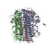
| ||||||||
|---|---|---|---|---|---|---|---|---|---|
| 1 |
| ||||||||
| Unit cell |
|
-Components
-Protein , 1 types, 3 molecules ABC
| #1: Protein | Mass: 42752.516 Da / Num. of mol.: 3 Source method: isolated from a genetically manipulated source Source: (gene. exp.) Streptomyces peucetius subsp. caesius ATCC 27952 (bacteria)Gene: CGZ69_03210 / Production host: References: UniProt: A0A2D3U3Z1, inulin fructotransferase (DFA-I-forming) |
|---|
-Non-polymers , 6 types, 603 molecules 


| #2: Chemical | ChemComp-GOL /  Mass: 92.094 Da / Num. of mol.: 18 / Source method: obtained synthetically / Formula: C3H8O3 Mass: 92.094 Da / Num. of mol.: 18 / Source method: obtained synthetically / Formula: C3H8O3#3: Chemical | ChemComp-EDO /  Mass: 62.068 Da / Num. of mol.: 22 / Source method: isolated from a natural source / Formula: C2H6O2 Mass: 62.068 Da / Num. of mol.: 22 / Source method: isolated from a natural source / Formula: C2H6O2#4: Chemical |  Mass: 238.278 Da / Num. of mol.: 2 / Source method: obtained synthetically / Formula: C10H22O6 / Comment: precipitant*YM Mass: 238.278 Da / Num. of mol.: 2 / Source method: obtained synthetically / Formula: C10H22O6 / Comment: precipitant*YM#5: Chemical | ChemComp-PEG /  Mass: 106.120 Da / Num. of mol.: 5 / Source method: obtained synthetically / Formula: C4H10O3 Mass: 106.120 Da / Num. of mol.: 5 / Source method: obtained synthetically / Formula: C4H10O3#6: Chemical |  Mass: 79.980 Da / Num. of mol.: 3 / Source method: obtained synthetically / Formula: HO3P Mass: 79.980 Da / Num. of mol.: 3 / Source method: obtained synthetically / Formula: HO3P#7: Water | ChemComp-HOH / | Mass: 18.015 Da / Num. of mol.: 553 / Source method: isolated from a natural source / Formula: H2O |
|---|
-Details
| Has ligand of interest | N |
|---|---|
| Has protein modification | N |
-Experimental details
-Experiment
| Experiment | Method: X-RAY DIFFRACTION / Number of used crystals: 1 |
|---|
- Sample preparation
| Crystal | Density Matthews: 2.59 Å3/Da / Density % sol: 53.62 % |
|---|---|
| Crystal grow | Temperature: 289.15 K / Method: vapor diffusion, sitting drop / pH: 6.5 Details: 0.1 M MES monohydrate, 12% Polyethylene glycol 20,000 |
-Data collection
| Diffraction | Mean temperature: 100 K / Serial crystal experiment: N |
|---|---|
| Diffraction source | Source: SYNCHROTRON / Site: SSRF / Beamline: BL18U1 / Wavelength: 0.97915 Å |
| Detector | Type: DECTRIS PILATUS3 6M / Detector: PIXEL / Date: Dec 24, 2020 |
| Radiation | Protocol: SINGLE WAVELENGTH / Monochromatic (M) / Laue (L): M / Scattering type: x-ray |
| Radiation wavelength | Wavelength: 0.97915 Å / Relative weight: 1 |
| Reflection | Resolution: 1.69→47.81 Å / Num. obs: 147518 / % possible obs: 99 % / Redundancy: 13.3 % / CC1/2: 0.999 / Rmerge(I) obs: 0.085 / Rrim(I) all: 0.088 / Net I/σ(I): 22.8 |
| Reflection shell | Resolution: 1.69→1.75 Å / Rmerge(I) obs: 0.36 / Num. unique obs: 14488 / CC1/2: 0.969 / Rrim(I) all: 0.374 |
- Processing
| Software |
| |||||||||||||||||||||||||||||||||||||||||||||||||||||||||||||||||||||||||||||||||||||||||||||||||||||||||
|---|---|---|---|---|---|---|---|---|---|---|---|---|---|---|---|---|---|---|---|---|---|---|---|---|---|---|---|---|---|---|---|---|---|---|---|---|---|---|---|---|---|---|---|---|---|---|---|---|---|---|---|---|---|---|---|---|---|---|---|---|---|---|---|---|---|---|---|---|---|---|---|---|---|---|---|---|---|---|---|---|---|---|---|---|---|---|---|---|---|---|---|---|---|---|---|---|---|---|---|---|---|---|---|---|---|---|
| Refinement | Method to determine structure: MOLECULAR REPLACEMENT / Resolution: 1.69→47.81 Å / SU ML: 0.1 / Cross valid method: THROUGHOUT / σ(F): 1.36 / Phase error: 12.99 / Stereochemistry target values: ML
| |||||||||||||||||||||||||||||||||||||||||||||||||||||||||||||||||||||||||||||||||||||||||||||||||||||||||
| Solvent computation | Shrinkage radii: 0.9 Å / VDW probe radii: 1.1 Å / Solvent model: FLAT BULK SOLVENT MODEL | |||||||||||||||||||||||||||||||||||||||||||||||||||||||||||||||||||||||||||||||||||||||||||||||||||||||||
| Refinement step | Cycle: LAST / Resolution: 1.69→47.81 Å
| |||||||||||||||||||||||||||||||||||||||||||||||||||||||||||||||||||||||||||||||||||||||||||||||||||||||||
| Refine LS restraints |
| |||||||||||||||||||||||||||||||||||||||||||||||||||||||||||||||||||||||||||||||||||||||||||||||||||||||||
| LS refinement shell |
|
