- PDB-8hml: Co-crystal structure of the C terminal DNA binding domain of Sacc... -
+
Open data
ID or keywords:
Loading...
-
Basic information
Entry
Database: PDB / ID: 8hml
Title
Co-crystal structure of the C terminal DNA binding domain of Saccharopolyspora erythraea GlnR in complex with its conserved promoter DNA in 2.95 Angstrom resolution
Components
DNA (5'-D(*AP*CP*GP*TP*AP*AP*CP*AP*TP*CP*GP*CP*GP*GP*TP*AP*AP*CP*AP*C)-3')
DNA (5'-D(*GP*TP*GP*TP*TP*AP*CP*CP*GP*CP*GP*AP*TP*GP*TP*TP*AP*CP*GP*T)-3')
National Natural Science Foundation of China (NSFC)
81903526
China
Citation
Journal: Proc Natl Acad Sci U S A / Year: 2023 Title: Structural insights into the transcription activation mechanism of the global regulator GlnR from actinobacteria. Authors: Jing Shi / Zhenzhen Feng / Juncao Xu / Fangfang Li / Yuqiong Zhang / Aijia Wen / Fulin Wang / Qian Song / Lu Wang / Hong Cui / Shujuan Tong / Peiying Chen / Yejin Zhu / Guoping Zhao / Shuang ...Authors: Jing Shi / Zhenzhen Feng / Juncao Xu / Fangfang Li / Yuqiong Zhang / Aijia Wen / Fulin Wang / Qian Song / Lu Wang / Hong Cui / Shujuan Tong / Peiying Chen / Yejin Zhu / Guoping Zhao / Shuang Wang / Yu Feng / Wei Lin / Abstract: In actinobacteria, an OmpR/PhoB subfamily protein called GlnR acts as an orphan response regulator and globally coordinates the expression of genes responsible for nitrogen, carbon, and phosphate ...In actinobacteria, an OmpR/PhoB subfamily protein called GlnR acts as an orphan response regulator and globally coordinates the expression of genes responsible for nitrogen, carbon, and phosphate metabolism in actinobacteria. Although many researchers have attempted to elucidate the mechanisms of GlnR-dependent transcription activation, progress is impeded by lacking of an overall structure of GlnR-dependent transcription activation complex (GlnR-TAC). Here, we report a co-crystal structure of the C-terminal DNA-binding domain of GlnR (GlnR_DBD) in complex with its regulatory -element DNA and a cryo-EM structure of GlnR-TAC which comprises RNA polymerase, GlnR, and a promoter containing four well-characterized conserved GlnR binding sites. These structures illustrate how four GlnR protomers coordinate to engage promoter DNA in a head-to-tail manner, with four N-terminal receiver domains of GlnR (GlnR-RECs) bridging GlnR_DBDs and the RNAP core enzyme. Structural analysis also unravels that GlnR-TAC is stabilized by complex protein-protein interactions between GlnR and the conserved β flap, σR4, αCTD, and αNTD domains of RNAP, which are further confirmed by our biochemical assays. Taken together, these results reveal a global transcription activation mechanism for the master regulator GlnR and other OmpR/PhoB subfamily proteins and present a unique mode of bacterial transcription regulation.
History
Deposition
Dec 4, 2022
Deposition site: PDBJ / Processing site: PDBJ
Revision 1.0
Jun 7, 2023
Provider: repository / Type: Initial release
Revision 1.1
May 29, 2024
Group: Data collection / Category: chem_comp_atom / chem_comp_bond
A: DNA-binding response OmpR family regulator B: DNA-binding response OmpR family regulator D: DNA (5'-D(*AP*CP*GP*TP*AP*AP*CP*AP*TP*CP*GP*CP*GP*GP*TP*AP*AP*CP*AP*C)-3') C: DNA (5'-D(*GP*TP*GP*TP*TP*AP*CP*CP*GP*CP*GP*AP*TP*GP*TP*TP*AP*CP*GP*T)-3')
In the structure databanks used in Yorodumi, some data are registered as the other names, "COVID-19 virus" and "2019-nCoV". Here are the details of the virus and the list of structure data.
Jan 31, 2019. EMDB accession codes are about to change! (news from PDBe EMDB page)
EMDB accession codes are about to change! (news from PDBe EMDB page)
The allocation of 4 digits for EMDB accession codes will soon come to an end. Whilst these codes will remain in use, new EMDB accession codes will include an additional digit and will expand incrementally as the available range of codes is exhausted. The current 4-digit format prefixed with “EMD-” (i.e. EMD-XXXX) will advance to a 5-digit format (i.e. EMD-XXXXX), and so on. It is currently estimated that the 4-digit codes will be depleted around Spring 2019, at which point the 5-digit format will come into force.
The EM Navigator/Yorodumi systems omit the EMD- prefix.
Related info.:Q: What is EMD? / ID/Accession-code notation in Yorodumi/EM Navigator
Yorodumi is a browser for structure data from EMDB, PDB, SASBDB, etc.
This page is also the successor to EM Navigator detail page, and also detail information page/front-end page for Omokage search.
The word "yorodu" (or yorozu) is an old Japanese word meaning "ten thousand". "mi" (miru) is to see.
Related info.:EMDB / PDB / SASBDB / Comparison of 3 databanks / Yorodumi Search / Aug 31, 2016. New EM Navigator & Yorodumi / Yorodumi Papers / Jmol/JSmol / Function and homology information / Changes in new EM Navigator and Yorodumi
 Movie
Movie Controller

 Yorodumi
 Open data
 Basic information
 Components
 Keywords
 Function and homology information
 Saccharopolyspora erythraea NRRL 2338 (bacteria)
 X-RAY DIFFRACTION /
  Authors
 China, 1items
  Citation
 Structure visualization
 Downloads & links
 Other downloads

 PDBj
PDBj


 Assembly
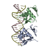
 Mass: 18.015 Da / Num. of mol.: 15 / Source method: isolated from a natural source / Formula: H2O
 Sample preparation
 Processing