 Movie
Movie Controller

[English] 日本語
 Yorodumi
- PDB-8hm2: Crystal structure of human ubiquitin-like protein from bacteroide... -
+ Open data

- Basic information
| Entry | Database: PDB / ID: 8hm2 | ||||||
|---|---|---|---|---|---|---|---|
| Title | Crystal structure of human ubiquitin-like protein from bacteroides fragilis c terminal cysteine mutant | ||||||
 Components Components | Putative ubiquitin | ||||||
 Keywords Keywords | CELL CYCLE / ubiqutin like protein | ||||||
| Function / homology | : / Ubiquitin domain / Ubiquitin family / Ubiquitin homologues / Ubiquitin domain profile. / Ubiquitin-like domain / Ubiquitin-like domain superfamily / Ubiquitin Function and homology information Function and homology information | ||||||
| Biological species |  Bacteroides fragilis (bacteria) Bacteroides fragilis (bacteria) | ||||||
| Method |  X-RAY DIFFRACTION / SYNCHROTRON / MOLECULAR REPLACEMENT / Resolution: 1.34 Å X-RAY DIFFRACTION / SYNCHROTRON / MOLECULAR REPLACEMENT / Resolution: 1.34 Å | ||||||
 Authors Authors | Tong, M. / Chen, Z. / Gao, X. | ||||||
| Funding support |  China, 1items China, 1items
| ||||||
 Citation Citation | Journal: Nat Microbiol / Year: 2024 Title: Bacteroides fragilis ubiquitin homologue drives intraspecies bacterial competition in the gut microbiome. Authors: Jiang, K. / Li, W. / Tong, M. / Xu, J. / Chen, Z. / Yang, Y. / Zang, Y. / Jiao, X. / Liu, C. / Lim, B. / Jiang, X. / Wang, J. / Wu, D. / Wang, M. / Liu, S.J. / Shao, F. / Gao, X. | ||||||
| History |
|
- Structure visualization
| Structure viewer | Molecule: MolmilJmol/JSmol |
|---|
- Downloads & links
-Download
| PDBx/mmCIF format | 8hm2.cif.gz | 48.1 KB | Display | PDBx/mmCIF format |
|---|---|---|---|---|
| PDB format | pdb8hm2.ent.gz | 32 KB | Display | PDB format |
| PDBx/mmJSON format | 8hm2.json.gz | Tree view | PDBx/mmJSON format | |
| Others |  Other downloads Other downloads |
-Validation report
| Arichive directory | https://data.pdbj.org/pub/pdb/validation_reports/hm/8hm2ftp://data.pdbj.org/pub/pdb/validation_reports/hm/8hm2 | HTTPS FTP |
|---|
-Related structure data
| Related structure data | 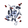 8hm1C  8hm3C  8hm4C  1ubqS S: Starting model for refinement C: citing same article ( |
|---|---|
| Similar structure data |
-Links
 PDBj
PDBj- Assembly
| Deposited unit | 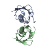
| ||||||||||||
|---|---|---|---|---|---|---|---|---|---|---|---|---|---|
| 1 | 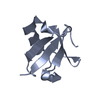
| ||||||||||||
| 2 | 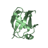
| ||||||||||||
| Unit cell |
| ||||||||||||
| Components on special symmetry positions |
|
-Components
| #1: Protein | Mass: 8828.024 Da / Num. of mol.: 2 / Mutation: C70V, C76G Source method: isolated from a genetically manipulated source Source: (gene. exp.) Bacteroides fragilis (bacteria) / Gene: ubb, BF9343_3779 / Production host: #2: Water | ChemComp-HOH / |  Mass: 18.015 Da / Num. of mol.: 137 / Source method: isolated from a natural source / Formula: H2O Mass: 18.015 Da / Num. of mol.: 137 / Source method: isolated from a natural source / Formula: H2O |
|---|
-Experimental details
-Experiment
| Experiment | Method: X-RAY DIFFRACTION / Number of used crystals: 1 |
|---|
- Sample preparation
| Crystal | Density Matthews: 1.89 Å3/Da / Density % sol: 34.99 % |
|---|---|
| Crystal grow | Temperature: 291 K / Method: vapor diffusion, hanging drop / pH: 2.8 / Details: 0.1M citric acid pH 2.8, 1.6 M ammonium sulfate |
-Data collection
| Diffraction | Mean temperature: 100 K / Serial crystal experiment: N |
|---|---|
| Diffraction source | Source: SYNCHROTRON / Site: SSRF / Beamline: BL02U1 / Wavelength: 0.97918 Å |
| Detector | Type: DECTRIS EIGER2 S 9M / Detector: PIXEL / Date: Nov 20, 2022 |
| Radiation | Protocol: SINGLE WAVELENGTH / Monochromatic (M) / Laue (L): M / Scattering type: x-ray |
| Radiation wavelength | Wavelength: 0.97918 Å / Relative weight: 1 |
| Reflection | Resolution: 1.34→29.07 Å / Num. obs: 30343 / % possible obs: 96.76 % / Redundancy: 20 % / Biso Wilson estimate: 15.96 Å2 / CC1/2: 0.994 / Net I/σ(I): 13.5 |
| Reflection shell | Resolution: 1.34→1.388 Å / Num. unique obs: 2397 / CC1/2: 0.697 |
- Processing
| Software |
| ||||||||||||||||||||||||||||||||||||||||||||||||||||||||||||||||||||||||||||||||||||
|---|---|---|---|---|---|---|---|---|---|---|---|---|---|---|---|---|---|---|---|---|---|---|---|---|---|---|---|---|---|---|---|---|---|---|---|---|---|---|---|---|---|---|---|---|---|---|---|---|---|---|---|---|---|---|---|---|---|---|---|---|---|---|---|---|---|---|---|---|---|---|---|---|---|---|---|---|---|---|---|---|---|---|---|---|---|
| Refinement | Method to determine structure: MOLECULAR REPLACEMENT Starting model: 1UBQ Resolution: 1.34→29.07 Å / SU ML: 0.2324 / Cross valid method: FREE R-VALUE / σ(F): 1.36 / Phase error: 29.6364 Stereochemistry target values: GeoStd + Monomer Library + CDL v1.2
| ||||||||||||||||||||||||||||||||||||||||||||||||||||||||||||||||||||||||||||||||||||
| Solvent computation | Shrinkage radii: 0.9 Å / VDW probe radii: 1.11 Å / Solvent model: FLAT BULK SOLVENT MODEL | ||||||||||||||||||||||||||||||||||||||||||||||||||||||||||||||||||||||||||||||||||||
| Displacement parameters | Biso mean: 22.45 Å2 | ||||||||||||||||||||||||||||||||||||||||||||||||||||||||||||||||||||||||||||||||||||
| Refinement step | Cycle: LAST / Resolution: 1.34→29.07 Å
| ||||||||||||||||||||||||||||||||||||||||||||||||||||||||||||||||||||||||||||||||||||
| Refine LS restraints |
| ||||||||||||||||||||||||||||||||||||||||||||||||||||||||||||||||||||||||||||||||||||
| LS refinement shell |
|
