[日本語] English
- PDB-8hk7: Structure of PKD2-F604P (Polycystin-2, TRPP2) with ML-SA1 -

+
データを開く


IDまたはキーワード:

読み込み中...

-
基本情報

登録情報
データベース: PDB / ID: 8hk7
タイトルStructure of PKD2-F604P (Polycystin-2, TRPP2) with ML-SA1
要素Polycystin-2
キーワードMEMBRANE PROTEIN
機能・相同性
機能・相同性情報


detection of nodal flow / metanephric smooth muscle tissue development / metanephric cortex development / metanephric cortical collecting duct development / metanephric distal tubule development / polycystin complex / mesonephric tubule development / mesonephric duct development / metanephric part of ureteric bud development / determination of liver left/right asymmetry ...detection of nodal flow / metanephric smooth muscle tissue development / metanephric cortex development / metanephric cortical collecting duct development / metanephric distal tubule development / polycystin complex / mesonephric tubule development / mesonephric duct development / metanephric part of ureteric bud development / determination of liver left/right asymmetry / renal tubule morphogenesis / metanephric ascending thin limb development / HLH domain binding / basal cortex / metanephric mesenchyme development / metanephric S-shaped body morphogenesis / renal artery morphogenesis / positive regulation of inositol 1,4,5-trisphosphate-sensitive calcium-release channel activity / migrasome / cilium organization / VxPx cargo-targeting to cilium / centrosome duplication / detection of mechanical stimulus / calcium-induced calcium release activity / regulation of calcium ion import / cation channel complex / muscle alpha-actinin binding / placenta blood vessel development / voltage-gated monoatomic ion channel activity / cellular response to hydrostatic pressure / outward rectifier potassium channel activity / cellular response to fluid shear stress / voltage-gated monoatomic cation channel activity / cellular response to osmotic stress / non-motile cilium / actinin binding / motile cilium / transcription regulator inhibitor activity / determination of left/right symmetry / aorta development / inorganic cation transmembrane transport / neural tube development / ciliary membrane / voltage-gated sodium channel activity / protein heterotetramerization / embryonic placenta development / branching involved in ureteric bud morphogenesis / negative regulation of G1/S transition of mitotic cell cycle / spinal cord development / heart looping / negative regulation of ryanodine-sensitive calcium-release channel activity / voltage-gated potassium channel activity / cytoplasmic side of endoplasmic reticulum membrane / cell surface receptor signaling pathway via JAK-STAT / potassium channel activity / sodium ion transmembrane transport / voltage-gated calcium channel activity / monoatomic cation channel activity / basal plasma membrane / cellular response to cAMP / release of sequestered calcium ion into cytosol / potassium ion transmembrane transport / cytoskeletal protein binding / cellular response to calcium ion / ciliary basal body / liver development / establishment of localization in cell / lumenal side of endoplasmic reticulum membrane / phosphoprotein binding / calcium ion transmembrane transport / protein tetramerization / cellular response to reactive oxygen species / cytoplasmic vesicle membrane / cilium / mitotic spindle / Wnt signaling pathway / intracellular calcium ion homeostasis / calcium ion transport / positive regulation of nitric oxide biosynthetic process / cell-cell junction / lamellipodium / ATPase binding / heart development / regulation of cell population proliferation / positive regulation of cytosolic calcium ion concentration / basolateral plasma membrane / protein homotetramerization / transmembrane transporter binding / cell surface receptor signaling pathway / regulation of cell cycle / negative regulation of cell population proliferation / signaling receptor binding / calcium ion binding / endoplasmic reticulum membrane / positive regulation of gene expression / Golgi apparatus / endoplasmic reticulum / positive regulation of transcription by RNA polymerase II / protein homodimerization activity / extracellular exosome
類似検索 - 分子機能
: / Ferredoxin I 4Fe-4S cluster domain / Polycystin domain / Polycystin domain / Polycystic kidney disease type 2 protein / Polycystin cation channel, PKD1/PKD2 / Polycystin cation channel / Voltage-dependent channel domain superfamily / EF-hand calcium-binding domain profile. / EF-hand domain / EF-hand domain pair
類似検索 - ドメイン・相同性
Chem-AQV / Polycystin-2
類似検索 - 構成要素
生物種Homo sapiens (ヒト)
手法電子顕微鏡法 / 単粒子再構成法 / クライオ電子顕微鏡法 / 解像度: 3 Å
データ登録者Chen, M.Y. / Su, Q. / Wang, Z.F. / Yu, Y.
資金援助 中国, 1件
組織認可番号
National Natural Science Foundation of China (NSFC)31930059 中国
引用ジャーナル: Proc Natl Acad Sci U S A / : 2024
タイトル: Molecular and structural basis of the dual regulation of the polycystin-2 ion channel by small-molecule ligands.
著者: Zhifei Wang / Mengying Chen / Qiang Su / Tiago D C Morais / Yan Wang / Elianna Nazginov / Akhilraj R Pillai / Feng Qian / Yigong Shi / Yong Yu /
要旨: Mutations in the gene, which encodes the polycystin-2 (PC2, also called TRPP2) protein, lead to autosomal dominant polycystic kidney disease (ADPKD). As a member of the transient receptor potential ...Mutations in the gene, which encodes the polycystin-2 (PC2, also called TRPP2) protein, lead to autosomal dominant polycystic kidney disease (ADPKD). As a member of the transient receptor potential (TRP) channel superfamily, PC2 functions as a non-selective cation channel. The activation and regulation of the PC2 channel are largely unknown, and direct binding of small-molecule ligands to this channel has not been reported. In this work, we found that most known small-molecule agonists of the mucolipin TRP (TRPML) channels inhibit the activity of the PC2_F604P, a gain-of-function mutant of the PC2 channel. However, two of them, ML-SA1 and SF-51, have dual regulatory effects, with low concentration further activating PC2_F604P, and high concentration leading to inactivation of the channel. With two cryo-electron microscopy (cryo-EM) structures, a molecular docking model, and mutagenesis results, we identified two distinct binding sites of ML-SA1 in PC2_F604P that are responsible for activation and inactivation, respectively. These results provide structural and functional insights into how ligands regulate PC2 channel function through unusual mechanisms and may help design compounds that are more efficient and specific in regulating the PC2 channel and potentially also for ADPKD treatment.
履歴
登録2022年11月25日登録サイト: PDBJ / 処理サイト: RCSB
改定 1.02024年3月27日Provider: repository / タイプ: Initial release

-
構造の表示

構造ビューア分子:
MolmilJmol/JSmol

ダウンロードとリンク

-
集合体

登録構造単位
A: Polycystin-2
B: Polycystin-2
C: Polycystin-2
D: Polycystin-2
ヘテロ分子


分子量 (理論値)分子数
合計 (水以外)268,26421
ポリマ-264,1194
非ポリマー4,14417
00
1


  • 登録構造と同一
  • 登録者が定義した集合体
  • 根拠: 電子顕微鏡法
タイプ名称対称操作
identity operation1_5551

-
要素

#1: タンパク質
Polycystin-2 / PC2 / Autosomal dominant polycystic kidney disease type II protein / Polycystic kidney disease 2 ...PC2 / Autosomal dominant polycystic kidney disease type II protein / Polycystic kidney disease 2 protein / Polycystwin / R48321 / Transient receptor potential cation channel subfamily P member 2


分子量: 66029.828 Da / 分子数: 4 / 由来タイプ: 組換発現 / 由来: (組換発現) Homo sapiens (ヒト) / 遺伝子: PKD2, TRPP2 / 発現宿主: Homo sapiens (ヒト) / 参照: UniProt: Q13563
#2: 糖
ChemComp-NAG / 2-acetamido-2-deoxy-beta-D-glucopyranose / N-acetyl-beta-D-glucosamine / 2-acetamido-2-deoxy-beta-D-glucose / 2-acetamido-2-deoxy-D-glucose / 2-acetamido-2-deoxy-glucose / N-ACETYL-D-GLUCOSAMINE / N-アセチル-β-D-グルコサミン


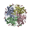
タイプ: D-saccharide, beta linking / 分子量: 221.208 Da / 分子数: 12 / 由来タイプ: 合成 / : C8H15NO6
識別子タイププログラム
DGlcpNAcbCONDENSED IUPAC CARBOHYDRATE SYMBOLGMML 1.0
N-acetyl-b-D-glucopyranosamineCOMMON NAMEGMML 1.0
b-D-GlcpNAcIUPAC CARBOHYDRATE SYMBOLPDB-CARE 1.0
GlcNAcSNFG CARBOHYDRATE SYMBOLGMML 1.0
#3: 化合物 ChemComp-CA / CALCIUM ION / カルシウムジカチオン


分子量: 40.078 Da / 分子数: 1 / 由来タイプ: 合成 / : Ca / タイプ: SUBJECT OF INVESTIGATION
#4: 化合物
ChemComp-AQV / 2-{2-oxo-2-[(4S)-2,2,4-trimethyl-3,4-dihydroquinolin-1(2H)-yl]ethyl}-1H-isoindole-1,3(2H)-dione


分子量: 362.422 Da / 分子数: 4 / 由来タイプ: 合成 / : C22H22N2O3 / タイプ: SUBJECT OF INVESTIGATION
研究の焦点であるリガンドがあるかY

-
実験情報

-
実験

実験手法: 電子顕微鏡法
EM実験試料の集合状態: PARTICLE / 3次元再構成法: 単粒子再構成法

-
試料調製

構成要素名称: Structure of PKD2-F604P (Polycystin-2, TRPP2) with ML-SA1
タイプ: COMPLEX / Entity ID: #1 / 由来: RECOMBINANT
由来(天然)生物種: Homo (哺乳類)
由来(組換発現)生物種: Homo (哺乳類)
緩衝液pH: 7.5
試料包埋: NO / シャドウイング: NO / 染色: NO / 凍結: YES
急速凍結凍結剤: ETHANE

-
電子顕微鏡撮影

実験機器
モデル: Titan Krios / 画像提供: FEI Company
顕微鏡モデル: FEI TITAN KRIOS
電子銃電子線源: FIELD EMISSION GUN / 加速電圧: 300 kV / 照射モード: FLOOD BEAM
電子レンズモード: BRIGHT FIELD / 最大 デフォーカス(公称値): 2000 nm / 最小 デフォーカス(公称値): 1400 nm
撮影電子線照射量: 50 e/Å2 / フィルム・検出器のモデル: GATAN K3 (6k x 4k)

-
解析

CTF補正タイプ: NONE
3次元再構成解像度: 3 Å / 解像度の算出法: FSC 0.143 CUT-OFF / 粒子像の数: 163172 / 対称性のタイプ: POINT
拘束条件
Refine-IDタイプDev ideal
ELECTRON MICROSCOPYf_bond_d0.00715316
ELECTRON MICROSCOPYf_angle_d0.64720836
ELECTRON MICROSCOPYf_dihedral_angle_d26.9452056
ELECTRON MICROSCOPYf_chiral_restr0.0432380
ELECTRON MICROSCOPYf_plane_restr0.0042588

+
万見について

-
お知らせ

-
2022年2月9日: EMDBエントリの付随情報ファイルのフォーマットが新しくなりました

EMDBエントリの付随情報ファイルのフォーマットが新しくなりました

  • EMDBのヘッダファイルのバージョン3が、公式のフォーマットとなりました。
  • これまでは公式だったバージョン1.9は、アーカイブから削除されます。

関連情報:EMDBヘッダ

外部リンク:wwPDBはEMDBデータモデルのバージョン3へ移行します

-
2020年8月12日: 新型コロナ情報

新型コロナ情報

URL: https://pdbjlc1.pdbj.org/emnavi/covid19.php

新ページ: EM Navigatorに新型コロナウイルスの特設ページを開設しました。

関連情報:Covid-19情報 / 2020年3月5日: 新型コロナウイルスの構造データ

+
2020年3月5日: 新型コロナウイルスの構造データ

新型コロナウイルスの構造データ

関連情報:万見生物種 / 2020年8月12日: 新型コロナ情報

外部リンク:COVID-19特集ページ - PDBj / 今月の分子2020年2月:コロナウイルスプロテーアーゼ

+
2019年1月31日: EMDBのIDの桁数の変更

EMDBのIDの桁数の変更

  • EMDBエントリに付与されているアクセスコード(EMDB-ID)は4桁の数字(例、EMD-1234)でしたが、間もなく枯渇します。これまでの4桁のID番号は4桁のまま変更されませんが、4桁の数字を使い切った後に発行されるIDは5桁以上の数字(例、EMD-12345)になります。5桁のIDは2019年の春頃から発行される見通しです。
  • EM Navigator/万見では、接頭語「EMD-」は省略されています。

関連情報:Q: 「EMD」とは何ですか? / 万見/EM NavigatorにおけるID/アクセスコードの表記

外部リンク:EMDB Accession Codes are Changing Soon! / PDBjへお問い合わせ

+
2017年7月12日: PDB大規模アップデート

PDB大規模アップデート

  • 新バージョンのPDBx/mmCIF辞書形式に基づくデータがリリースされました。
  • 今回の更新はバージョン番号が4から5になる大規模なもので、全エントリデータの書き換えが行われる「Remediation」というアップデートに該当します。
  • このバージョンアップで、電子顕微鏡の実験手法に関する多くの項目の書式が改定されました(例:em_softwareなど)。
  • EM NavigatorとYorodumiでも、この改定に基づいた表示内容になります。

外部リンク:wwPDB Remediation / OneDepデータ基準に準拠した、より強化された内容のモデル構造ファイルが、PDBアーカイブで公開されました。

-
万見 (Yorodumi)

幾万の構造データを、幾万の視点から

  • 万見(Yorodumi)は、EMDB/PDB/SASBDBなどの構造データを閲覧するためのページです。
  • EM Navigatorの詳細ページの後継、Omokage検索のフロントエンドも兼ねています。

関連情報:EMDB / PDB / SASBDB / 3つのデータバンクの比較 / 万見検索 / 2016年8月31日: 新しいEM Navigatorと万見 / 万見文献 / Jmol/JSmol / 機能・相同性情報 / 新しいEM Navigatorと万見の変更点

他の情報も見る