 Movie
Movie Controller


+ Open data

- Basic information
| Entry | Database: PDB / ID: 8his | |||||||||||||||||||||||||||||||||||||||||||||||||
|---|---|---|---|---|---|---|---|---|---|---|---|---|---|---|---|---|---|---|---|---|---|---|---|---|---|---|---|---|---|---|---|---|---|---|---|---|---|---|---|---|---|---|---|---|---|---|---|---|---|---|
| Title | Crystal structure of DNA decamer containing GuNA[Me,tBu] | |||||||||||||||||||||||||||||||||||||||||||||||||
 Components Components | DNA (5'-D(* Keywords KeywordsDNA / OLIGONUCLEOTIDE / MODIFIED BASE | Function / homology | CACODYLIC ACID / DNA |  Function and homology information Function and homology informationBiological species | synthetic construct (others) | Method |  X-RAY DIFFRACTION / MOLECULAR REPLACEMENT / molecular replacement / Resolution: 2.01 Å X-RAY DIFFRACTION / MOLECULAR REPLACEMENT / molecular replacement / Resolution: 2.01 Å  Authors AuthorsAoyama, H. / Obika, S. / Yamaguchi, T. | Funding support | |  Japan, 8items Japan, 8items
 CitationJournal: Nucleic Acids Res. / Year: 2023 CitationJournal: Nucleic Acids Res. / Year: 2023Title: Mechanism of the extremely high duplex-forming ability of oligonucleotides modified with N-tert-butylguanidine- or N-tert-butyl-N'- methylguanidine-bridged nucleic acids. Authors: Yamaguchi, T. / Horie, N. / Aoyama, H. / Kumagai, S. / Obika, S. History |
|
- Structure visualization
| Structure viewer | Molecule: MolmilJmol/JSmol |
|---|
- Downloads & links
-Download
| PDBx/mmCIF format | 8his.cif.gz | 26.1 KB | Display | PDBx/mmCIF format |
|---|---|---|---|---|
| PDB format | pdb8his.ent.gz | 14.8 KB | Display | PDB format |
| PDBx/mmJSON format | 8his.json.gz | Tree view | PDBx/mmJSON format | |
| Others |  Other downloads Other downloads |
-Validation report
| Arichive directory | https://data.pdbj.org/pub/pdb/validation_reports/hi/8hisftp://data.pdbj.org/pub/pdb/validation_reports/hi/8his | HTTPS FTP |
|---|
-Related structure data
| Related structure data |  8hu5C 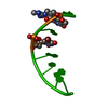 8i50C 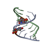 1i5wS S: Starting model for refinement C: citing same article ( |
|---|---|
| Similar structure data |
-Links
 PDBj
PDBj

- Assembly
| Deposited unit | 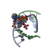
| ||||||||
|---|---|---|---|---|---|---|---|---|---|
| 1 |
| ||||||||
| Unit cell |
|
-Components
| #1: DNA chain | Mass: 3184.204 Da / Num. of mol.: 2 / Source method: obtained synthetically / Source: (synth.) synthetic construct (others) #2: Chemical | ChemComp-CAD / | 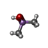  Mass: 137.997 Da / Num. of mol.: 1 / Source method: obtained synthetically / Formula: C2H7AsO2 Mass: 137.997 Da / Num. of mol.: 1 / Source method: obtained synthetically / Formula: C2H7AsO2#3: Water | ChemComp-HOH / |  Mass: 18.015 Da / Num. of mol.: 91 / Source method: isolated from a natural source / Formula: H2O Mass: 18.015 Da / Num. of mol.: 91 / Source method: isolated from a natural source / Formula: H2OHas ligand of interest | Y | |
|---|
-Experimental details
-Experiment
| Experiment | Method: X-RAY DIFFRACTION / Number of used crystals: 1 |
|---|
- Sample preparation
| Crystal | Density Matthews: 1.99 Å3/Da / Density % sol: 38.22 % |
|---|---|
| Crystal grow | Temperature: 293.15 K / Method: vapor diffusion, hanging drop Details: 10% v/v (+/-)-2-Methyl-2,4-pentanediol, 0.040M Sodium cacodylate trihydrate pH 6.0, 0.012M Spermine tetrahydrochloride, 0.080M Potassium chloride, 0.020M Barium chloride |
-Data collection
| Diffraction | Mean temperature: 100 K / Serial crystal experiment: N |
|---|---|
| Diffraction source | Source: ROTATING ANODE / Type: RIGAKU MICROMAX-007 HF / Wavelength: 1.5418 Å |
| Detector | Type: RIGAKU RAXIS IV++ / Detector: IMAGE PLATE / Date: Mar 26, 2019 |
| Radiation | Protocol: SINGLE WAVELENGTH / Monochromatic (M) / Laue (L): M / Scattering type: x-ray |
| Radiation wavelength | Wavelength: 1.5418 Å / Relative weight: 1 |
| Reflection | Resolution: 2.01→20.13 Å / Num. obs: 6474 / % possible obs: 99.7 % / Redundancy: 6.5 % / CC1/2: 0.999 / Rmerge(I) obs: 0.042 / Rpim(I) all: 0.018 / Rrim(I) all: 0.046 / Χ2: 0.94 / Net I/σ(I): 55.1 |
| Reflection shell | Resolution: 2.01→2.07 Å / % possible obs: 99.4 % / Redundancy: 6.4 % / Rmerge(I) obs: 0.055 / Num. measured all: 1890 / Num. unique obs: 294 / CC1/2: 0.998 / Rpim(I) all: 0.023 / Rrim(I) all: 0.06 / Χ2: 0.83 / Net I/σ(I) obs: 43.8 |
-Phasing
| Phasing | Method: molecular replacement | |||||||||
|---|---|---|---|---|---|---|---|---|---|---|
| Phasing MR | Model details: Phaser MODE: MR_AUTO
|
- Processing
| Software |
| ||||||||||||||||||||||||||||||||||||||||||
|---|---|---|---|---|---|---|---|---|---|---|---|---|---|---|---|---|---|---|---|---|---|---|---|---|---|---|---|---|---|---|---|---|---|---|---|---|---|---|---|---|---|---|---|
| Refinement | Method to determine structure: MOLECULAR REPLACEMENT Starting model: 1I5W Resolution: 2.01→20.13 Å / SU ML: 0.19 / Cross valid method: THROUGHOUT / σ(F): 1 / Phase error: 21.63 / Stereochemistry target values: ML
| ||||||||||||||||||||||||||||||||||||||||||
| Solvent computation | Shrinkage radii: 0.9 Å / VDW probe radii: 1.1 Å / Solvent model: FLAT BULK SOLVENT MODEL | ||||||||||||||||||||||||||||||||||||||||||
| Refinement step | Cycle: LAST / Resolution: 2.01→20.13 Å
| ||||||||||||||||||||||||||||||||||||||||||
| Refine LS restraints |
| ||||||||||||||||||||||||||||||||||||||||||
| LS refinement shell |
|
