 Movie
Movie Controller


+ Open data

- Basic information
| Entry | Database: PDB / ID: 8hgu | ||||||
|---|---|---|---|---|---|---|---|
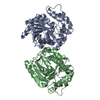
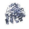
| Title | Epoxide hydrolase from Bosea sp. PAMC 26642 | ||||||
 Components Components | Alpha/beta hydrolase | ||||||
 Keywords Keywords | HYDROLASE | ||||||
| Biological species |  Bosea sp. PAMC 26642 (bacteria) Bosea sp. PAMC 26642 (bacteria) | ||||||
| Method |  X-RAY DIFFRACTION / SYNCHROTRON / MOLECULAR REPLACEMENT / Resolution: 1.94 Å X-RAY DIFFRACTION / SYNCHROTRON / MOLECULAR REPLACEMENT / Resolution: 1.94 Å | ||||||
 Authors Authors | Lee, M.J. / Hwang, J. / Do, H. / Lee, J.H. | ||||||
| Funding support |  Korea, Republic Of, 1items Korea, Republic Of, 1items
| ||||||
 Citation Citation | Journal: Int.J.Biol.Macromol. / Year: 2024 Title: Structural insights into the distinct substrate preferences of two bacterial epoxide hydrolases. Authors: Hwang, J. / Lee, M.J. / Lee, S.G. / Do, H. / Lee, J.H. | ||||||
| History |
|
- Structure visualization
| Structure viewer | Molecule:  MolmilJmol/JSmol MolmilJmol/JSmol |
|---|
- Downloads & links
-Download
| PDBx/mmCIF format | 8hgu.cif.gz | 250 KB | Display | PDBx/mmCIF format |
|---|---|---|---|---|
| PDB format | pdb8hgu.ent.gz | 197.4 KB | Display | PDB format |
| PDBx/mmJSON format | 8hgu.json.gz | Tree view | PDBx/mmJSON format | |
| Others |  Other downloads Other downloads |
-Validation report
| Summary document | 8hgu_validation.pdf.gz | 439.1 KB | Display | wwPDB validaton report |
|---|---|---|---|---|
| Full document | 8hgu_full_validation.pdf.gz | 441.6 KB | Display | |
| Data in XML | 8hgu_validation.xml.gz | 45.8 KB | Display | |
| Data in CIF | 8hgu_validation.cif.gz | 67.6 KB | Display | |
| Arichive directory | https://data.pdbj.org/pub/pdb/validation_reports/hg/8hguftp://data.pdbj.org/pub/pdb/validation_reports/hg/8hgu | HTTPS FTP |
-Related structure data
-Links
 PDBj
PDBj- Assembly
| Deposited unit | 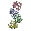
| ||||||||||||
|---|---|---|---|---|---|---|---|---|---|---|---|---|---|
| 1 | 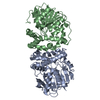
| ||||||||||||
| 2 | 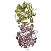
| ||||||||||||
| Unit cell |
|
-Components
| #1: Protein | Mass: 33256.266 Da / Num. of mol.: 4 Source method: isolated from a genetically manipulated source Source: (gene. exp.) Bosea sp. PAMC 26642 (bacteria) / Gene: AXW83_17570 / Production host: #2: Water | ChemComp-HOH / |  Mass: 18.015 Da / Num. of mol.: 644 / Source method: isolated from a natural source / Formula: H2O Mass: 18.015 Da / Num. of mol.: 644 / Source method: isolated from a natural source / Formula: H2O |
|---|
-Experimental details
-Experiment
| Experiment | Method: X-RAY DIFFRACTION / Number of used crystals: 1 |
|---|
- Sample preparation
| Crystal | Density Matthews: 2.69 Å3/Da / Density % sol: 54.36 % |
|---|---|
| Crystal grow | Temperature: 296.15 K / Method: vapor diffusion, sitting drop Details: 1 M Ammonium formate, 0.1 M Sodium acetate pH 5.0, 8 % w/v PGA (Na+ form, LM) |
-Data collection
| Diffraction | Mean temperature: 100 K / Serial crystal experiment: N |
|---|---|
| Diffraction source | Source: SYNCHROTRON / Site: PAL/PLS / Beamline: 7A (6B, 6C1) / Wavelength: 0.9793 Å |
| Detector | Type: ADSC QUANTUM 270 / Detector: CCD / Date: Apr 27, 2022 |
| Radiation | Protocol: SINGLE WAVELENGTH / Monochromatic (M) / Laue (L): M / Scattering type: x-ray |
| Radiation wavelength | Wavelength: 0.9793 Å / Relative weight: 1 |
| Reflection | Resolution: 1.94→50 Å / Num. obs: 101095 / % possible obs: 97.6 % / Redundancy: 4.4 % / Biso Wilson estimate: 19.34 Å2 / Rmerge(I) obs: 0.11 / Net I/σ(I): 29.96 |
| Reflection shell | Resolution: 1.95→1.98 Å / Redundancy: 2.7 % / Rmerge(I) obs: 0.41 / Mean I/σ(I) obs: 4.23 / Num. unique obs: 4384 / % possible all: 85.3 |
- Processing
| Software |
| ||||||||||||||||||||||||||||||||||||||||||||||||||||||||||||||||||||||||||||||||||||||||||||||||||||||||||||||||||||||||||||||||||||||||||||||||||||||||||||||||||||||||||||||||||||||||||||||||||||||||||||||||||
|---|---|---|---|---|---|---|---|---|---|---|---|---|---|---|---|---|---|---|---|---|---|---|---|---|---|---|---|---|---|---|---|---|---|---|---|---|---|---|---|---|---|---|---|---|---|---|---|---|---|---|---|---|---|---|---|---|---|---|---|---|---|---|---|---|---|---|---|---|---|---|---|---|---|---|---|---|---|---|---|---|---|---|---|---|---|---|---|---|---|---|---|---|---|---|---|---|---|---|---|---|---|---|---|---|---|---|---|---|---|---|---|---|---|---|---|---|---|---|---|---|---|---|---|---|---|---|---|---|---|---|---|---|---|---|---|---|---|---|---|---|---|---|---|---|---|---|---|---|---|---|---|---|---|---|---|---|---|---|---|---|---|---|---|---|---|---|---|---|---|---|---|---|---|---|---|---|---|---|---|---|---|---|---|---|---|---|---|---|---|---|---|---|---|---|---|---|---|---|---|---|---|---|---|---|---|---|---|---|---|---|---|
| Refinement | Method to determine structure: MOLECULAR REPLACEMENT Starting model: 1EHY Resolution: 1.94→30.2 Å / SU ML: 0.1886 / Cross valid method: FREE R-VALUE / σ(F): 1.45 / Phase error: 19.951 Stereochemistry target values: GeoStd + Monomer Library + CDL v1.2
| ||||||||||||||||||||||||||||||||||||||||||||||||||||||||||||||||||||||||||||||||||||||||||||||||||||||||||||||||||||||||||||||||||||||||||||||||||||||||||||||||||||||||||||||||||||||||||||||||||||||||||||||||||
| Solvent computation | Shrinkage radii: 0.9 Å / VDW probe radii: 1.1 Å / Solvent model: FLAT BULK SOLVENT MODEL | ||||||||||||||||||||||||||||||||||||||||||||||||||||||||||||||||||||||||||||||||||||||||||||||||||||||||||||||||||||||||||||||||||||||||||||||||||||||||||||||||||||||||||||||||||||||||||||||||||||||||||||||||||
| Displacement parameters | Biso mean: 21.8 Å2 | ||||||||||||||||||||||||||||||||||||||||||||||||||||||||||||||||||||||||||||||||||||||||||||||||||||||||||||||||||||||||||||||||||||||||||||||||||||||||||||||||||||||||||||||||||||||||||||||||||||||||||||||||||
| Refinement step | Cycle: LAST / Resolution: 1.94→30.2 Å
| ||||||||||||||||||||||||||||||||||||||||||||||||||||||||||||||||||||||||||||||||||||||||||||||||||||||||||||||||||||||||||||||||||||||||||||||||||||||||||||||||||||||||||||||||||||||||||||||||||||||||||||||||||
| Refine LS restraints |
| ||||||||||||||||||||||||||||||||||||||||||||||||||||||||||||||||||||||||||||||||||||||||||||||||||||||||||||||||||||||||||||||||||||||||||||||||||||||||||||||||||||||||||||||||||||||||||||||||||||||||||||||||||
| LS refinement shell |
|
