 Movie
Movie Controller

[English] 日本語
 Yorodumi
- PDB-8hge: Crystal structure of the CYP153A mutant V456A from Marinobacter a... -
+ Open data

- Basic information
| Entry | Database: PDB / ID: 8hge | ||||||
|---|---|---|---|---|---|---|---|
| Title | Crystal structure of the CYP153A mutant V456A from Marinobacter aquaeolei in complex with 12-hydroxydodecanoic acid | ||||||
 Components Components | Cytochrome P450 | ||||||
 Keywords Keywords | OXIDOREDUCTASE / CYP153A / Complex | ||||||
| Function / homology |  Function and homology information Function and homology informationoxidoreductase activity, acting on paired donors, with incorporation or reduction of molecular oxygen / monooxygenase activity / iron ion binding / heme binding Similarity search - Function | ||||||
| Biological species |  Marinobacter nauticus (bacteria) Marinobacter nauticus (bacteria) | ||||||
| Method |  X-RAY DIFFRACTION / SYNCHROTRON / MOLECULAR REPLACEMENT / Resolution: 1.53 Å X-RAY DIFFRACTION / SYNCHROTRON / MOLECULAR REPLACEMENT / Resolution: 1.53 Å | ||||||
 Authors Authors | Jiang, Y. / Cong, Z. | ||||||
| Funding support |  China, 1items China, 1items
| ||||||
 Citation Citation | Journal: To Be Published Title: Crystal structure of the CYP153A mutant V456A from Marinobacter aquaeolei in complex with 12-hydroxydodecanoic acid Authors: Jiang, Y. / Cong, Z. | ||||||
| History |
|
- Structure visualization
| Structure viewer | Molecule: MolmilJmol/JSmol |
|---|
- Downloads & links
-Download
| PDBx/mmCIF format | 8hge.cif.gz | 197.7 KB | Display | PDBx/mmCIF format |
|---|---|---|---|---|
| PDB format | pdb8hge.ent.gz | 150.9 KB | Display | PDB format |
| PDBx/mmJSON format | 8hge.json.gz | Tree view | PDBx/mmJSON format | |
| Others |  Other downloads Other downloads |
-Validation report
| Arichive directory | https://data.pdbj.org/pub/pdb/validation_reports/hg/8hgeftp://data.pdbj.org/pub/pdb/validation_reports/hg/8hge | HTTPS FTP |
|---|
-Related structure data
| Related structure data |  5fygS S: Starting model for refinement |
|---|---|
| Similar structure data |
-Links
 PDBj
PDBj
- Assembly
| Deposited unit | 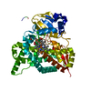
| ||||||||
|---|---|---|---|---|---|---|---|---|---|
| 1 |
| ||||||||
| Unit cell |
|
-Components
| #1: Protein | Mass: 55250.988 Da / Num. of mol.: 1 / Mutation: V456A Source method: isolated from a genetically manipulated source Source: (gene. exp.) Marinobacter nauticus (bacteria) / Gene: DET51_1164 / Production host: |
|---|---|
| #2: Chemical | ChemComp-HEM /   Mass: 616.487 Da / Num. of mol.: 1 / Source method: obtained synthetically / Formula: C34H32FeN4O4 / Feature type: SUBJECT OF INVESTIGATION Mass: 616.487 Da / Num. of mol.: 1 / Source method: obtained synthetically / Formula: C34H32FeN4O4 / Feature type: SUBJECT OF INVESTIGATION |
| #3: Chemical | ChemComp-12H /   Mass: 216.317 Da / Num. of mol.: 1 / Source method: obtained synthetically / Formula: C12H24O3 / Feature type: SUBJECT OF INVESTIGATION Mass: 216.317 Da / Num. of mol.: 1 / Source method: obtained synthetically / Formula: C12H24O3 / Feature type: SUBJECT OF INVESTIGATION |
| #4: Water | ChemComp-HOH /  Mass: 18.015 Da / Num. of mol.: 549 / Source method: isolated from a natural source / Formula: H2O Mass: 18.015 Da / Num. of mol.: 549 / Source method: isolated from a natural source / Formula: H2O |
| Has ligand of interest | Y |
| Has protein modification | N |
-Experimental details
-Experiment
| Experiment | Method: X-RAY DIFFRACTION / Number of used crystals: 1 |
|---|
- Sample preparation
| Crystal | Density Matthews: 2.04 Å3/Da / Density % sol: 39.64 % |
|---|---|
| Crystal grow | Temperature: 291 K / Method: vapor diffusion, hanging drop / pH: 7 / Details: 0.2 M Li2SO4, 0.1 M HEPES 7.0, 26% PEG3350 |
-Data collection
| Diffraction | Mean temperature: 100 K / Serial crystal experiment: N |
|---|---|
| Diffraction source | Source: SYNCHROTRON / Site: SSRF / Beamline: BL19U1 / Wavelength: 0.979 Å |
| Detector | Type: DECTRIS PILATUS3 6M / Detector: PIXEL / Date: Oct 14, 2022 |
| Radiation | Protocol: SINGLE WAVELENGTH / Monochromatic (M) / Laue (L): M / Scattering type: x-ray |
| Radiation wavelength | Wavelength: 0.979 Å / Relative weight: 1 |
| Reflection | Resolution: 1.53→50 Å / Num. obs: 63827 / % possible obs: 95.6 % / Redundancy: 3.3 % / CC1/2: 0.998 / Rmerge(I) obs: 0.048 / Rpim(I) all: 0.031 / Rrim(I) all: 0.057 / Χ2: 0.894 / Net I/σ(I): 19.9 |
| Reflection shell | Resolution: 1.53→1.56 Å / Redundancy: 2.7 % / Rmerge(I) obs: 0.132 / Num. unique obs: 2949 / CC1/2: 0.964 / Rpim(I) all: 0.097 / Rrim(I) all: 0.165 / Χ2: 0.794 / % possible all: 87.9 |
- Processing
| Software |
| |||||||||||||||||||||||||||||||||||||||||||||||||||||||||||||||||||||||||||||||||||||||||||||||||||||||||
|---|---|---|---|---|---|---|---|---|---|---|---|---|---|---|---|---|---|---|---|---|---|---|---|---|---|---|---|---|---|---|---|---|---|---|---|---|---|---|---|---|---|---|---|---|---|---|---|---|---|---|---|---|---|---|---|---|---|---|---|---|---|---|---|---|---|---|---|---|---|---|---|---|---|---|---|---|---|---|---|---|---|---|---|---|---|---|---|---|---|---|---|---|---|---|---|---|---|---|---|---|---|---|---|---|---|---|
| Refinement | Method to determine structure: MOLECULAR REPLACEMENT Starting model: 5FYG Resolution: 1.53→31.54 Å / SU ML: 0.1 / Cross valid method: THROUGHOUT / σ(F): 1.48 / Phase error: 17.28 / Stereochemistry target values: ML
| |||||||||||||||||||||||||||||||||||||||||||||||||||||||||||||||||||||||||||||||||||||||||||||||||||||||||
| Solvent computation | Shrinkage radii: 0.9 Å / VDW probe radii: 1.11 Å / Solvent model: FLAT BULK SOLVENT MODEL | |||||||||||||||||||||||||||||||||||||||||||||||||||||||||||||||||||||||||||||||||||||||||||||||||||||||||
| Refinement step | Cycle: LAST / Resolution: 1.53→31.54 Å
| |||||||||||||||||||||||||||||||||||||||||||||||||||||||||||||||||||||||||||||||||||||||||||||||||||||||||
| Refine LS restraints |
| |||||||||||||||||||||||||||||||||||||||||||||||||||||||||||||||||||||||||||||||||||||||||||||||||||||||||
| LS refinement shell |
|
