 Movie
Movie Controller


+ Open data

- Basic information
| Entry | Database: PDB / ID: 8hen | ||||||
|---|---|---|---|---|---|---|---|
| Title | Crystal structure of CTSB in complex with 212-148 | ||||||
 Components Components | Cathepsin B | ||||||
 Keywords Keywords | ANTIVIRAL PROTEIN/INHIBITOR / inhibitor / antiviral / protease / ANTIVIRAL PROTEIN / ANTIVIRAL PROTEIN-INHIBITOR complex | ||||||
| Function / homology |  Function and homology information Function and homology informationcathepsin B / thyroid hormone generation / endolysosome lumen / cellular response to thyroid hormone stimulus / Trafficking and processing of endosomal TLR / proteoglycan binding / Assembly of collagen fibrils and other multimeric structures / Collagen degradation / collagen catabolic process / collagen binding ...cathepsin B / thyroid hormone generation / endolysosome lumen / cellular response to thyroid hormone stimulus / Trafficking and processing of endosomal TLR / proteoglycan binding / Assembly of collagen fibrils and other multimeric structures / Collagen degradation / collagen catabolic process / collagen binding / epithelial cell differentiation / cysteine-type peptidase activity / MHC class II antigen presentation / : / Degradation of CDH1 / melanosome / peptidase activity / extracellular matrix / regulation of apoptotic process / ficolin-1-rich granule lumen / lysosome / apical plasma membrane / external side of plasma membrane / cysteine-type endopeptidase activity / Neutrophil degranulation / symbiont entry into host cell / perinuclear region of cytoplasm / proteolysis / : / extracellular exosome / extracellular region Similarity search - Function | ||||||
| Biological species |  Homo sapiens (human) Homo sapiens (human) | ||||||
| Method |  X-RAY DIFFRACTION / SYNCHROTRON / MOLECULAR REPLACEMENT / Resolution: 1.95 Å X-RAY DIFFRACTION / SYNCHROTRON / MOLECULAR REPLACEMENT / Resolution: 1.95 Å | ||||||
 Authors Authors | Wang, H. / Li, D. / Sun, L. / Yang, H. | ||||||
| Funding support |  China, 1items China, 1items
| ||||||
 Citation Citation | Journal: Nat Commun / Year: 2023 Title: Structure-based discovery of dual pathway inhibitors for SARS-CoV-2 entry. Authors: Wang, H. / Yang, Q. / Liu, X. / Xu, Z. / Shao, M. / Li, D. / Duan, Y. / Tang, J. / Yu, X. / Zhang, Y. / Hao, A. / Wang, Y. / Chen, J. / Zhu, C. / Guddat, L. / Chen, H. / Zhang, L. / Chen, X. ...Authors: Wang, H. / Yang, Q. / Liu, X. / Xu, Z. / Shao, M. / Li, D. / Duan, Y. / Tang, J. / Yu, X. / Zhang, Y. / Hao, A. / Wang, Y. / Chen, J. / Zhu, C. / Guddat, L. / Chen, H. / Zhang, L. / Chen, X. / Jiang, B. / Sun, L. / Rao, Z. / Yang, H. | ||||||
| History |
|
- Structure visualization
| Structure viewer | Molecule: MolmilJmol/JSmol |
|---|
- Downloads & links
-Download
| PDBx/mmCIF format | 8hen.cif.gz | 70.4 KB | Display | PDBx/mmCIF format |
|---|---|---|---|---|
| PDB format | pdb8hen.ent.gz | 48.3 KB | Display | PDB format |
| PDBx/mmJSON format | 8hen.json.gz | Tree view | PDBx/mmJSON format | |
| Others |  Other downloads Other downloads |
-Validation report
| Arichive directory | https://data.pdbj.org/pub/pdb/validation_reports/he/8henftp://data.pdbj.org/pub/pdb/validation_reports/he/8hen | HTTPS FTP |
|---|
-Related structure data
| Related structure data |  7xydC  7y0eC  7y0fC  8hd8C  8he9C  8heiC  8hetC  8hfvC 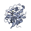 6ay2S S: Starting model for refinement C: citing same article ( |
|---|---|
| Similar structure data |
-Links
 PDBj
PDBj


- Assembly
| Deposited unit | 
| ||||||||
|---|---|---|---|---|---|---|---|---|---|
| 1 |
| ||||||||
| Unit cell |
|
-Components
| #1: Protein | Mass: 28200.402 Da / Num. of mol.: 1 Source method: isolated from a genetically manipulated source Source: (gene. exp.) Homo sapiens (human) / Gene: CTSB, CPSB / Production host:  | ||||||||||
|---|---|---|---|---|---|---|---|---|---|---|---|
| #2: Chemical |   Mass: 92.094 Da / Num. of mol.: 2 / Source method: isolated from a natural source / Formula: C3H8O3 Mass: 92.094 Da / Num. of mol.: 2 / Source method: isolated from a natural source / Formula: C3H8O3#3: Chemical | ChemComp-DMS / |   Mass: 78.133 Da / Num. of mol.: 1 / Source method: obtained synthetically / Formula: C2H6OS / Comment: DMSO, precipitant*YM Mass: 78.133 Da / Num. of mol.: 1 / Source method: obtained synthetically / Formula: C2H6OS / Comment: DMSO, precipitant*YM#4: Chemical | ChemComp-L4F / |   Mass: 767.936 Da / Num. of mol.: 1 / Source method: obtained synthetically / Formula: C41H49N7O6S / Feature type: SUBJECT OF INVESTIGATION Mass: 767.936 Da / Num. of mol.: 1 / Source method: obtained synthetically / Formula: C41H49N7O6S / Feature type: SUBJECT OF INVESTIGATION#5: Water | ChemComp-HOH / |  Mass: 18.015 Da / Num. of mol.: 139 / Source method: isolated from a natural source / Formula: H2O Mass: 18.015 Da / Num. of mol.: 139 / Source method: isolated from a natural source / Formula: H2OHas ligand of interest | Y | Has protein modification | Y | |
-Experimental details
-Experiment
| Experiment | Method: X-RAY DIFFRACTION / Number of used crystals: 1 |
|---|
- Sample preparation
| Crystal | Density Matthews: 2.14 Å3/Da / Density % sol: 42.55 % |
|---|---|
| Crystal grow | Temperature: 293 K / Method: vapor diffusion, hanging drop Details: 0.2 M Magnesium chloride hexahydrate, 0.1 M NaAc pH 5.2, 28% w/v PEG 3350 |
-Data collection
| Diffraction | Mean temperature: 100 K / Serial crystal experiment: N | ||||||||||||||||||||||||||||||||||||||||||||||||||||||||||||
|---|---|---|---|---|---|---|---|---|---|---|---|---|---|---|---|---|---|---|---|---|---|---|---|---|---|---|---|---|---|---|---|---|---|---|---|---|---|---|---|---|---|---|---|---|---|---|---|---|---|---|---|---|---|---|---|---|---|---|---|---|---|
| Diffraction source | Source: SYNCHROTRON / Site: SSRF / Beamline: BL10U2 / Wavelength: 0.979 Å | ||||||||||||||||||||||||||||||||||||||||||||||||||||||||||||
| Detector | Type: DECTRIS EIGER X 16M / Detector: PIXEL / Date: Aug 17, 2022 | ||||||||||||||||||||||||||||||||||||||||||||||||||||||||||||
| Radiation | Protocol: SINGLE WAVELENGTH / Monochromatic (M) / Laue (L): M / Scattering type: x-ray | ||||||||||||||||||||||||||||||||||||||||||||||||||||||||||||
| Radiation wavelength | Wavelength: 0.979 Å / Relative weight: 1 | ||||||||||||||||||||||||||||||||||||||||||||||||||||||||||||
| Reflection | Resolution: 1.95→37.47 Å / Num. obs: 18398 / % possible obs: 99.5 % / Redundancy: 6.09 % / CC1/2: 0.996 / Rmerge(I) obs: 0.148 / Rrim(I) all: 0.162 / Net I/σ(I): 12.19 | ||||||||||||||||||||||||||||||||||||||||||||||||||||||||||||
| Reflection shell |
|
- Processing
| Software |
| ||||||||||||||||||||||||||||||||||||||||||||||||||||||||||||||||||||||||||||||||||||||||||||||||||
|---|---|---|---|---|---|---|---|---|---|---|---|---|---|---|---|---|---|---|---|---|---|---|---|---|---|---|---|---|---|---|---|---|---|---|---|---|---|---|---|---|---|---|---|---|---|---|---|---|---|---|---|---|---|---|---|---|---|---|---|---|---|---|---|---|---|---|---|---|---|---|---|---|---|---|---|---|---|---|---|---|---|---|---|---|---|---|---|---|---|---|---|---|---|---|---|---|---|---|---|
| Refinement | Method to determine structure: MOLECULAR REPLACEMENT Starting model: 6AY2 Resolution: 1.95→37.47 Å / SU ML: 0.24 / Cross valid method: THROUGHOUT / σ(F): 0.88 / Phase error: 22.97 / Stereochemistry target values: ML
| ||||||||||||||||||||||||||||||||||||||||||||||||||||||||||||||||||||||||||||||||||||||||||||||||||
| Solvent computation | Shrinkage radii: 0.9 Å / VDW probe radii: 1.1 Å / Solvent model: FLAT BULK SOLVENT MODEL | ||||||||||||||||||||||||||||||||||||||||||||||||||||||||||||||||||||||||||||||||||||||||||||||||||
| Refinement step | Cycle: LAST / Resolution: 1.95→37.47 Å
| ||||||||||||||||||||||||||||||||||||||||||||||||||||||||||||||||||||||||||||||||||||||||||||||||||
| Refine LS restraints |
| ||||||||||||||||||||||||||||||||||||||||||||||||||||||||||||||||||||||||||||||||||||||||||||||||||
| LS refinement shell |
|
