 Movie
Movie Controller


+ Open data

- Basic information
| Entry | Database: PDB / ID: 8hcy | ||||||
|---|---|---|---|---|---|---|---|
| Title | Legionella glycosyltransferase | ||||||
 Components Components | Dot/Icm secretion system substrate | ||||||
 Keywords Keywords | TRANSFERASE / glucosyltransferase / upd-glucose | ||||||
| Function / homology | Glycosyltransferase family 88 / Glycosyltransferase family 88 / Dot/Icm secretion system substrate Function and homology information Function and homology information | ||||||
| Biological species |  Legionella (bacteria) Legionella (bacteria) | ||||||
| Method |  X-RAY DIFFRACTION / SYNCHROTRON / MOLECULAR REPLACEMENT / Resolution: 2.64 Å X-RAY DIFFRACTION / SYNCHROTRON / MOLECULAR REPLACEMENT / Resolution: 2.64 Å | ||||||
 Authors Authors | Chen, T.T. / Ouyang, S.Y. | ||||||
| Funding support |  China, 1items China, 1items
| ||||||
 Citation Citation | Journal: Small Struct / Year: 2023 Title: Structural Basis for the Action Mechanism of Legionella Glycosyltransferase. Authors: Chen, T.T. / Zheng, S.R. / Tian, L. / Lv, S.L. / Zhong, W.H. / Li, X.L. / Zhang, D.D. / Zheng, X.X. / Ouyang, S.Y. | ||||||
| History |
|
- Structure visualization
| Structure viewer | Molecule: MolmilJmol/JSmol |
|---|
- Downloads & links
-Download
| PDBx/mmCIF format | 8hcy.cif.gz | 836.1 KB | Display | PDBx/mmCIF format |
|---|---|---|---|---|
| PDB format | pdb8hcy.ent.gz | 648 KB | Display | PDB format |
| PDBx/mmJSON format | 8hcy.json.gz | Tree view | PDBx/mmJSON format | |
| Others |  Other downloads Other downloads |
-Validation report
| Arichive directory | https://data.pdbj.org/pub/pdb/validation_reports/hc/8hcyftp://data.pdbj.org/pub/pdb/validation_reports/hc/8hcy | HTTPS FTP |
|---|
-Related structure data
| Related structure data | 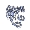 7xgvSC 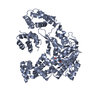 7xgxC S: Starting model for refinement C: citing same article ( |
|---|---|
| Similar structure data |
-Links
 PDBj
PDBj- Assembly
| Deposited unit | 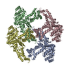
| ||||||||||||
|---|---|---|---|---|---|---|---|---|---|---|---|---|---|
| 1 | 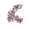
| ||||||||||||
| 2 | 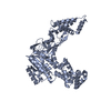
| ||||||||||||
| 3 | 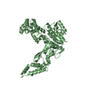
| ||||||||||||
| 4 | 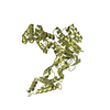
| ||||||||||||
| Unit cell |
|
-Components
| #1: Protein | Mass: 98615.453 Da / Num. of mol.: 4 Source method: isolated from a genetically manipulated source Source: (gene. exp.) Legionella (bacteria) / Gene: C3927_06670 / Production host: #2: Water | ChemComp-HOH / |  Mass: 18.015 Da / Num. of mol.: 392 / Source method: isolated from a natural source / Formula: H2O Mass: 18.015 Da / Num. of mol.: 392 / Source method: isolated from a natural source / Formula: H2O |
|---|
-Experimental details
-Experiment
| Experiment | Method: X-RAY DIFFRACTION / Number of used crystals: 1 |
|---|
- Sample preparation
| Crystal | Density Matthews: 1.58 Å3/Da / Density % sol: 21.96 % |
|---|---|
| Crystal grow | Temperature: 289 K / Method: vapor diffusion, sitting drop / Details: PEG |
-Data collection
| Diffraction | Mean temperature: 100 K / Serial crystal experiment: N |
|---|---|
| Diffraction source | Source: SYNCHROTRON / Site: SSRF / Beamline: BL17B1 / Wavelength: 0.9792 Å |
| Detector | Type: DECTRIS PILATUS 200K / Detector: PIXEL / Date: Jul 14, 2021 |
| Radiation | Protocol: SINGLE WAVELENGTH / Monochromatic (M) / Laue (L): M / Scattering type: x-ray |
| Radiation wavelength | Wavelength: 0.9792 Å / Relative weight: 1 |
| Reflection | Resolution: 2.64→29.31 Å / Num. obs: 71942 / % possible obs: 95.51 % / Redundancy: 6.7 % / Biso Wilson estimate: 28.23 Å2 / CC1/2: 0.971 / Rmerge(I) obs: 0.13 / Net I/σ(I): 4.07 |
| Reflection shell | Resolution: 2.64→2.73 Å / Num. unique obs: 68773 / CC1/2: 0.314 |
- Processing
| Software |
| |||||||||||||||||||||||||||||||||||||||||||||||||||||||||||||||||||||||||||||||||||||||||||||||||||||||||
|---|---|---|---|---|---|---|---|---|---|---|---|---|---|---|---|---|---|---|---|---|---|---|---|---|---|---|---|---|---|---|---|---|---|---|---|---|---|---|---|---|---|---|---|---|---|---|---|---|---|---|---|---|---|---|---|---|---|---|---|---|---|---|---|---|---|---|---|---|---|---|---|---|---|---|---|---|---|---|---|---|---|---|---|---|---|---|---|---|---|---|---|---|---|---|---|---|---|---|---|---|---|---|---|---|---|---|
| Refinement | Method to determine structure: MOLECULAR REPLACEMENT Starting model: 7XGV Resolution: 2.64→29.31 Å / SU ML: 0.2921 / Cross valid method: FREE R-VALUE / σ(F): 0.15 / Phase error: 27.563 Stereochemistry target values: GeoStd + Monomer Library + CDL v1.2
| |||||||||||||||||||||||||||||||||||||||||||||||||||||||||||||||||||||||||||||||||||||||||||||||||||||||||
| Solvent computation | Shrinkage radii: 0.9 Å / VDW probe radii: 1.11 Å / Solvent model: FLAT BULK SOLVENT MODEL | |||||||||||||||||||||||||||||||||||||||||||||||||||||||||||||||||||||||||||||||||||||||||||||||||||||||||
| Displacement parameters | Biso mean: 36.66 Å2 | |||||||||||||||||||||||||||||||||||||||||||||||||||||||||||||||||||||||||||||||||||||||||||||||||||||||||
| Refinement step | Cycle: LAST / Resolution: 2.64→29.31 Å
| |||||||||||||||||||||||||||||||||||||||||||||||||||||||||||||||||||||||||||||||||||||||||||||||||||||||||
| Refine LS restraints |
| |||||||||||||||||||||||||||||||||||||||||||||||||||||||||||||||||||||||||||||||||||||||||||||||||||||||||
| LS refinement shell |
|
