 Movie
Movie Controller


+ Open data

- Basic information
| Entry | Database: PDB / ID: 7xgx | ||||||
|---|---|---|---|---|---|---|---|
| Title | Glucosyltransferase | ||||||
 Components Components | Lgt2 | ||||||
 Keywords Keywords | TRANSFERASE / Glucosyltransferase / complex / bacterial effector | ||||||
| Function / homology | Glycosyltransferase family 88 / Glycosyltransferase family 88 / URIDINE-5'-DIPHOSPHATE-GLUCOSE / Glucosyltransferase Lgt2 Function and homology information Function and homology information | ||||||
| Biological species |  Legionella pneumophila subsp. pneumophila str. Philadelphia 1 (bacteria) Legionella pneumophila subsp. pneumophila str. Philadelphia 1 (bacteria) | ||||||
| Method |  X-RAY DIFFRACTION / SYNCHROTRON / MOLECULAR REPLACEMENT / Resolution: 2.39 Å X-RAY DIFFRACTION / SYNCHROTRON / MOLECULAR REPLACEMENT / Resolution: 2.39 Å | ||||||
 Authors Authors | Chen, T.T. / Ouyang, S.Y. | ||||||
| Funding support |  China, 1items China, 1items
| ||||||
 Citation Citation | Journal: Small Struct / Year: 2023 Title: Structural Basis for the Action Mechanism of Legionella Glycosyltransferase. Authors: Chen, T.T. / Zheng, S.R. / Tian, L. / Lv, S.L. / Zhong, W.H. / Li, X.L. / Zhang, D.D. / Zheng, X.X. / Ouyang, S.Y. | ||||||
| History |
|
- Structure visualization
| Structure viewer | Molecule: MolmilJmol/JSmol |
|---|
- Downloads & links
-Download
| PDBx/mmCIF format | 7xgx.cif.gz | 251.9 KB | Display | PDBx/mmCIF format |
|---|---|---|---|---|
| PDB format | pdb7xgx.ent.gz | 199.1 KB | Display | PDB format |
| PDBx/mmJSON format | 7xgx.json.gz | Tree view | PDBx/mmJSON format | |
| Others |  Other downloads Other downloads |
-Validation report
| Arichive directory | https://data.pdbj.org/pub/pdb/validation_reports/xg/7xgxftp://data.pdbj.org/pub/pdb/validation_reports/xg/7xgx | HTTPS FTP |
|---|
-Related structure data
| Related structure data | 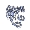 7xgvSC 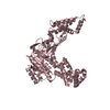 8hcyC S: Starting model for refinement C: citing same article ( |
|---|---|
| Similar structure data |
-Links
 PDBj
PDBj
- Assembly
| Deposited unit | 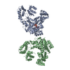
| ||||||||
|---|---|---|---|---|---|---|---|---|---|
| 1 | 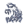
| ||||||||
| 2 | 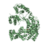
| ||||||||
| Unit cell |
|
-Components
| #1: Protein | Mass: 73831.320 Da / Num. of mol.: 2 Source method: isolated from a genetically manipulated source Source: (gene. exp.) Legionella pneumophila subsp. pneumophila str. Philadelphia 1 (bacteria)Strain: Philadelphia 1 / ATCC 33152 / DSM 7513 / Gene: C3927_14430 / Production host: #2: Chemical |   Mass: 566.302 Da / Num. of mol.: 2 / Source method: obtained synthetically / Formula: C15H24N2O17P2 / Feature type: SUBJECT OF INVESTIGATION Mass: 566.302 Da / Num. of mol.: 2 / Source method: obtained synthetically / Formula: C15H24N2O17P2 / Feature type: SUBJECT OF INVESTIGATION#3: Water | ChemComp-HOH / |  Mass: 18.015 Da / Num. of mol.: 120 / Source method: isolated from a natural source / Formula: H2O Mass: 18.015 Da / Num. of mol.: 120 / Source method: isolated from a natural source / Formula: H2OHas ligand of interest | Y | |
|---|
-Experimental details
-Experiment
| Experiment | Method: X-RAY DIFFRACTION / Number of used crystals: 1 |
|---|
- Sample preparation
| Crystal | Density Matthews: 2.44 Å3/Da / Density % sol: 49.62 % |
|---|---|
| Crystal grow | Temperature: 289 K / Method: vapor diffusion, hanging drop / Details: PEG3350, Calcium acetate hydrate |
-Data collection
| Diffraction | Mean temperature: 80 K / Serial crystal experiment: N |
|---|---|
| Diffraction source | Source: SYNCHROTRON / Site: SSRF / Beamline: BL17B1 / Wavelength: 0.979 Å |
| Detector | Type: APEX II CCD / Detector: CCD / Date: Dec 25, 2020 |
| Radiation | Protocol: SINGLE WAVELENGTH / Monochromatic (M) / Laue (L): M / Scattering type: x-ray |
| Radiation wavelength | Wavelength: 0.979 Å / Relative weight: 1 |
| Reflection | Resolution: 2.39→32.22 Å / Num. obs: 55366 / % possible obs: 95.28 % / Redundancy: 2 % / Biso Wilson estimate: 50.27 Å2 / CC1/2: 0.999 / Rmerge(I) obs: 0.02562 / Rpim(I) all: 0.02562 / Rrim(I) all: 0.03624 / Net I/σ(I): 14.72 |
| Reflection shell | Resolution: 2.39→2.475 Å / Rmerge(I) obs: 0.3477 / Mean I/σ(I) obs: 2.39 / Num. unique obs: 5597 / CC1/2: 0.867 / Rpim(I) all: 0.3477 / Rrim(I) all: 0.4917 |
- Processing
| Software |
| |||||||||||||||||||||||||||||||||||||||||||||||||||||||||||||||||||||||||||||||||||||||||||||||||||||||||
|---|---|---|---|---|---|---|---|---|---|---|---|---|---|---|---|---|---|---|---|---|---|---|---|---|---|---|---|---|---|---|---|---|---|---|---|---|---|---|---|---|---|---|---|---|---|---|---|---|---|---|---|---|---|---|---|---|---|---|---|---|---|---|---|---|---|---|---|---|---|---|---|---|---|---|---|---|---|---|---|---|---|---|---|---|---|---|---|---|---|---|---|---|---|---|---|---|---|---|---|---|---|---|---|---|---|---|
| Refinement | Method to determine structure: MOLECULAR REPLACEMENT Starting model: 7XGV Resolution: 2.39→32.22 Å / SU ML: 0.31 / Cross valid method: THROUGHOUT / σ(F): 0 / Phase error: 31.32 / Stereochemistry target values: ML
| |||||||||||||||||||||||||||||||||||||||||||||||||||||||||||||||||||||||||||||||||||||||||||||||||||||||||
| Solvent computation | Shrinkage radii: 0.9 Å / VDW probe radii: 1.11 Å / Solvent model: FLAT BULK SOLVENT MODEL | |||||||||||||||||||||||||||||||||||||||||||||||||||||||||||||||||||||||||||||||||||||||||||||||||||||||||
| Displacement parameters | Biso max: 221.13 Å2 / Biso mean: 65.5673 Å2 / Biso min: 30 Å2 | |||||||||||||||||||||||||||||||||||||||||||||||||||||||||||||||||||||||||||||||||||||||||||||||||||||||||
| Refinement step | Cycle: final / Resolution: 2.39→32.22 Å
| |||||||||||||||||||||||||||||||||||||||||||||||||||||||||||||||||||||||||||||||||||||||||||||||||||||||||
| LS refinement shell | Refine-ID: X-RAY DIFFRACTION / Rfactor Rfree error: 0 / Total num. of bins used: 14
|
