[English] 日本語
 Yorodumi
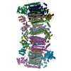
- PDB-8hcr: Cryo-EM structure of the Mycobacterium tuberculosis cytochrome bc... -
+ Open data
- Basic information
| Entry | Database: PDB / ID: 8hcr | |||||||||||||||||||||||||||||||||
|---|---|---|---|---|---|---|---|---|---|---|---|---|---|---|---|---|---|---|---|---|---|---|---|---|---|---|---|---|---|---|---|---|---|---|
| Title | Cryo-EM structure of the Mycobacterium tuberculosis cytochrome bcc:aa3 supercomplex and a novel inhibitor targeting subunit cytochrome cI | |||||||||||||||||||||||||||||||||
 Components Components |
| |||||||||||||||||||||||||||||||||
 Keywords Keywords | OXIDOREDUCTASE / Cytochrome bcc:aa3 oxidase / Mycobacterium tuberculosis | |||||||||||||||||||||||||||||||||
| Function / homology |  Function and homology information Function and homology informationaerobic electron transport chain / cytochrome-c oxidase / oxidative phosphorylation / quinol-cytochrome-c reductase / quinol-cytochrome-c reductase activity / cytochrome-c oxidase activity / electron transport coupled proton transport / oxidoreductase activity, acting on paired donors, with incorporation or reduction of molecular oxygen / aerobic respiration / respiratory electron transport chain ...aerobic electron transport chain / cytochrome-c oxidase / oxidative phosphorylation / quinol-cytochrome-c reductase / quinol-cytochrome-c reductase activity / cytochrome-c oxidase activity / electron transport coupled proton transport / oxidoreductase activity, acting on paired donors, with incorporation or reduction of molecular oxygen / aerobic respiration / respiratory electron transport chain / monooxygenase activity / 2 iron, 2 sulfur cluster binding / iron ion binding / heme binding / metal ion binding / membrane / plasma membrane Similarity search - Function | |||||||||||||||||||||||||||||||||
| Biological species |  Mycobacterium tuberculosis variant bovis BCG (bacteria) Mycobacterium tuberculosis variant bovis BCG (bacteria) | |||||||||||||||||||||||||||||||||
| Method | ELECTRON MICROSCOPY / electron tomography / cryo EM / Resolution: 4.5 Å | |||||||||||||||||||||||||||||||||
 Authors Authors | Mathiyazakan, V. / Gruber, G. | |||||||||||||||||||||||||||||||||
| Funding support |  Singapore, 1items Singapore, 1items
| |||||||||||||||||||||||||||||||||
 Citation Citation |  Journal: Antimicrob Agents Chemother / Year: 2023 Journal: Antimicrob Agents Chemother / Year: 2023Title: Cryo-Electron Microscopy Structure of the s Cytochrome : Supercomplex and a Novel Inhibitor Targeting Subunit Cytochrome I. Authors: Vikneswaran Mathiyazakan / Chui-Fann Wong / Amaravadhi Harikishore / Kevin Pethe / Gerhard Grüber /  Abstract: The mycobacterial cytochrome complex deserves the name "supercomplex" since it combines three cytochrome oxidases-cytochrome , cytochrome , and cytochrome -into one supramolecular machine and ...The mycobacterial cytochrome complex deserves the name "supercomplex" since it combines three cytochrome oxidases-cytochrome , cytochrome , and cytochrome -into one supramolecular machine and performs electron transfer for the reduction of oxygen to water and proton transport to generate the proton motive force for ATP synthesis. Thus, the complex represents a valid drug target for Mycobacterium tuberculosis infections. The production and purification of an entire M. tuberculosis cytochrome are fundamental for biochemical and structural characterization of this supercomplex, paving the way for new inhibitor targets and molecules. Here, we produced and purified the entire and active M. tuberculosis cyt- oxidase, as demonstrated by the different heme spectra and an oxygen consumption assay. The resolved M. tuberculosis cyt- cryo-electron microscopy structure reveals a dimer with its functional domains involved in electron, proton, oxygen transfer, and oxygen reduction. The structure shows the two cytochrome III head domains of the dimer, the counterpart of the soluble mitochondrial cytochrome , in a so-called "closed state," in which electrons are translocated from the to the domain. The structural and mechanistic insights provided the basis for a virtual screening campaign that identified a potent M. tuberculosis cyt- inhibitor, cyt1. cyt1 targets the mycobacterium-specific α3-helix of cytochrome I and interferes with oxygen consumption by interrupting electron translocation via the III head. The successful identification of a new cyt- inhibitor demonstrates the potential of a structure-mechanism-based approach for novel compound development. | |||||||||||||||||||||||||||||||||
| History |
|
- Structure visualization
| Structure viewer | Molecule:  Molmil Molmil Jmol/JSmol Jmol/JSmol |
|---|
- Downloads & links
- Download
| PDBx/mmCIF format |  8hcr.cif.gz 8hcr.cif.gz | 863.4 KB | Display |  PDBx/mmCIF format PDBx/mmCIF format |
|---|---|---|---|---|
| PDB format |  pdb8hcr.ent.gz pdb8hcr.ent.gz | 702.2 KB | Display |  PDB format PDB format |
| PDBx/mmJSON format |  8hcr.json.gz 8hcr.json.gz | Tree view |  PDBx/mmJSON format PDBx/mmJSON format | |
| Others |  Other downloads Other downloads |
-Validation report
| Arichive directory |  https://data.pdbj.org/pub/pdb/validation_reports/hc/8hcr https://data.pdbj.org/pub/pdb/validation_reports/hc/8hcr ftp://data.pdbj.org/pub/pdb/validation_reports/hc/8hcr ftp://data.pdbj.org/pub/pdb/validation_reports/hc/8hcr | HTTPS FTP |
|---|
-Related structure data
| Related structure data |  34664MC M: map data used to model this data C: citing same article ( |
|---|---|
| Similar structure data | Similarity search - Function & homology  F&H Search F&H Search |
- Links
Links
- Assembly
| Deposited unit | 
|
|---|---|
| 1 |
|
- Components
-Protein , 3 types, 6 molecules AMHTJV
| #1: Protein | Mass: 46976.465 Da / Num. of mol.: 2 Source method: isolated from a genetically manipulated source Source: (gene. exp.)  Mycobacterium tuberculosis variant bovis BCG (bacteria) Mycobacterium tuberculosis variant bovis BCG (bacteria)Gene: aioB, qcrA, ERS007663_00404, ERS007670_01335, ERS007679_00448, ERS007681_03213, ERS007703_01913, ERS007720_01586, ERS007722_02477, ERS007741_02707, SAMEA2683035_01305 Production host:  Mycobacterium tuberculosis variant bovis BCG (bacteria) Mycobacterium tuberculosis variant bovis BCG (bacteria)References: UniProt: A0A045GW18 #7: Protein | Mass: 14874.137 Da / Num. of mol.: 2 Source method: isolated from a genetically manipulated source Source: (gene. exp.)  Mycobacterium tuberculosis variant bovis BCG (bacteria) Mycobacterium tuberculosis variant bovis BCG (bacteria)Gene: RN06_2699 Production host:  Mycobacterium tuberculosis variant bovis BCG (bacteria) Mycobacterium tuberculosis variant bovis BCG (bacteria)References: UniProt: A0A0K2HXG0, cytochrome-c oxidase #9: Protein | Mass: 15982.042 Da / Num. of mol.: 2 Source method: isolated from a genetically manipulated source Source: (gene. exp.)  Mycobacterium tuberculosis variant bovis BCG (bacteria) Mycobacterium tuberculosis variant bovis BCG (bacteria)Gene: K60_025630 Production host:  Mycobacterium tuberculosis variant bovis BCG (bacteria) Mycobacterium tuberculosis variant bovis BCG (bacteria)References: UniProt: A0A7U4BVZ0 |
|---|
-Cytochrome bc1 complex cytochrome ... , 2 types, 4 molecules BNCO
| #2: Protein | Mass: 63925.984 Da / Num. of mol.: 2 Source method: isolated from a genetically manipulated source Source: (gene. exp.)  Mycobacterium tuberculosis variant bovis BCG (bacteria) Mycobacterium tuberculosis variant bovis BCG (bacteria)Gene: RN06_2696 Production host:  Mycobacterium tuberculosis variant bovis BCG (bacteria) Mycobacterium tuberculosis variant bovis BCG (bacteria)References: UniProt: A0A0K2HYC0, quinol-cytochrome-c reductase #3: Protein | Mass: 29170.404 Da / Num. of mol.: 2 Source method: isolated from a genetically manipulated source Source: (gene. exp.)  Mycobacterium tuberculosis variant bovis BCG (bacteria) Mycobacterium tuberculosis variant bovis BCG (bacteria)Gene: qcrC, Rv2194, MTCY190.05 Production host:  Mycobacterium tuberculosis variant bovis BCG (bacteria) Mycobacterium tuberculosis variant bovis BCG (bacteria)References: UniProt: P9WP35, quinol-cytochrome-c reductase |
|---|
-CYTOCHROME AA3 SUBUNIT ... , 2 types, 4 molecules EQIU
| #4: Protein | Mass: 40535.582 Da / Num. of mol.: 2 Source method: isolated from a genetically manipulated source Source: (gene. exp.)  Mycobacterium tuberculosis variant bovis BCG (bacteria) Mycobacterium tuberculosis variant bovis BCG (bacteria)Production host:  Mycobacterium tuberculosis variant bovis BCG (bacteria) Mycobacterium tuberculosis variant bovis BCG (bacteria)#8: Protein | Mass: 8408.646 Da / Num. of mol.: 2 Source method: isolated from a genetically manipulated source Source: (gene. exp.)  Mycobacterium tuberculosis variant bovis BCG (bacteria) Mycobacterium tuberculosis variant bovis BCG (bacteria)Production host:  Mycobacterium tuberculosis variant bovis BCG (bacteria) Mycobacterium tuberculosis variant bovis BCG (bacteria) |
|---|
-Probable cytochrome c oxidase subunit ... , 2 types, 4 molecules FRGS
| #5: Protein | Mass: 63722.289 Da / Num. of mol.: 2 Source method: isolated from a genetically manipulated source Source: (gene. exp.)  Mycobacterium tuberculosis variant bovis BCG (bacteria) Mycobacterium tuberculosis variant bovis BCG (bacteria)Gene: ctaD, Rv3043c, MTV012.58c Production host:  Mycobacterium tuberculosis variant bovis BCG (bacteria) Mycobacterium tuberculosis variant bovis BCG (bacteria)References: UniProt: P9WP71, cytochrome-c oxidase #6: Protein | Mass: 22435.021 Da / Num. of mol.: 2 Source method: isolated from a genetically manipulated source Source: (gene. exp.)  Mycobacterium tuberculosis variant bovis BCG (bacteria) Mycobacterium tuberculosis variant bovis BCG (bacteria)Gene: ctaE, Rv2193, MTCY190.04 Production host:  Mycobacterium tuberculosis variant bovis BCG (bacteria) Mycobacterium tuberculosis variant bovis BCG (bacteria)References: UniProt: P9WP67, cytochrome-c oxidase |
|---|
-Non-polymers , 4 types, 14 molecules 


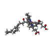
| #10: Chemical | | #11: Chemical | ChemComp-HEM / #12: Chemical | ChemComp-HEC / #13: Chemical | ChemComp-HEA / |
|---|
-Details
| Has ligand of interest | Y |
|---|---|
| Has protein modification | Y |
-Experimental details
-Experiment
| Experiment | Method: ELECTRON MICROSCOPY |
|---|---|
| EM experiment | Aggregation state: PARTICLE / 3D reconstruction method: electron tomography |
- Sample preparation
| Component | Name: Cytochrome bcc:aa3 supercomplex / Type: COMPLEX / Entity ID: #1-#9 / Source: RECOMBINANT |
|---|---|
| Molecular weight | Experimental value: NO |
| Source (natural) | Organism:  Mycobacterium tuberculosis variant bovis BCG (bacteria) Mycobacterium tuberculosis variant bovis BCG (bacteria) |
| Source (recombinant) | Organism:  Mycobacterium tuberculosis variant bovis BCG (bacteria) Mycobacterium tuberculosis variant bovis BCG (bacteria) |
| Buffer solution | pH: 7.4 |
| Specimen | Embedding applied: NO / Shadowing applied: NO / Staining applied: NO / Vitrification applied: YES |
| Vitrification | Cryogen name: ETHANE |
- Electron microscopy imaging
| Experimental equipment |  Model: Titan Krios / Image courtesy: FEI Company |
|---|---|
| Microscopy | Model: FEI TITAN KRIOS |
| Electron gun | Electron source:  FIELD EMISSION GUN / Accelerating voltage: 300 kV / Illumination mode: FLOOD BEAM FIELD EMISSION GUN / Accelerating voltage: 300 kV / Illumination mode: FLOOD BEAM |
| Electron lens | Mode: BRIGHT FIELD / Nominal defocus max: 2000 nm / Nominal defocus min: 1000 nm |
| Image recording | Electron dose: 40 e/Å2 / Detector mode: SUPER-RESOLUTION / Film or detector model: GATAN K2 SUMMIT (4k x 4k) |
- Processing
| Software | Name: PHENIX / Version: 1.20.1_4487: / Classification: refinement | ||||||||||||||||||||||||
|---|---|---|---|---|---|---|---|---|---|---|---|---|---|---|---|---|---|---|---|---|---|---|---|---|---|
| EM software | Name: PHENIX / Category: model refinement | ||||||||||||||||||||||||
| CTF correction | Type: PHASE FLIPPING AND AMPLITUDE CORRECTION | ||||||||||||||||||||||||
| 3D reconstruction | Resolution: 4.5 Å / Num. of particles: 100 | ||||||||||||||||||||||||
| Refine LS restraints |
|
 Movie
Movie Controller


 PDBj
PDBj


