 Movie
Movie Controller


+ Open data

- Basic information
| Entry | Database: PDB / ID: 8hbg | |||||||||||||||||||||||||||||||||
|---|---|---|---|---|---|---|---|---|---|---|---|---|---|---|---|---|---|---|---|---|---|---|---|---|---|---|---|---|---|---|---|---|---|---|
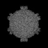
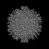
| Title | FMDV (A/TUR/14/98) in complex with M678F | |||||||||||||||||||||||||||||||||
 Components Components |
| |||||||||||||||||||||||||||||||||
 Keywords Keywords | VIRUS / FMDV / antibody | |||||||||||||||||||||||||||||||||
| Function / homology |  Function and homology information Function and homology informationT=pseudo3 icosahedral viral capsid / host cell cytoplasm / symbiont entry into host cell / virion attachment to host cell / structural molecule activity Similarity search - Function | |||||||||||||||||||||||||||||||||
| Biological species |   Foot-and-mouth disease virus A Foot-and-mouth disease virus A | |||||||||||||||||||||||||||||||||
| Method | ELECTRON MICROSCOPY / single particle reconstruction / cryo EM / Resolution: 3.6 Å | |||||||||||||||||||||||||||||||||
 Authors Authors | Li, H.Z. / Dong, H. / Liu, P. | |||||||||||||||||||||||||||||||||
| Funding support | 1items
| |||||||||||||||||||||||||||||||||
 Citation Citation | Journal: Nat Commun / Year: 2024 Title: Foot-and-mouth disease virus antigenic landscape and reduced immunogenicity elucidated in atomic detail. Authors: Haozhou Li / Pan Liu / Hu Dong / Aldo Dekker / Michiel M Harmsen / Huichen Guo / Xiangxi Wang / Shiqi Sun /   Abstract: Unlike most other picornaviruses, foot-and-mouth disease (FMD) intact virions (146S) dissociate easily into small pentameric subunits (12S). This causes a dramatically decreased immunogenicity by a ...Unlike most other picornaviruses, foot-and-mouth disease (FMD) intact virions (146S) dissociate easily into small pentameric subunits (12S). This causes a dramatically decreased immunogenicity by a mechanism that remains elusive. Here, we present the high-resolution structures of 12S (3.2 Å) and its immune complex of a single-domain antibody (VHH) targeting the particle interior (3.2 Å), as well as two 146S-specific VHHs complexed to distinct sites on the 146S capsid surface (3.6 Å and 2.9 Å). The antigenic landscape of 146S is depicted using 13 known FMD virus-antibody complexes. Comparison of the immunogenicity of 146S and 12S in pigs, focusing on the resulting antigenic sites and incorporating structural analysis, reveals that dissociation of 146S leads to structural alteration and destruction of multiple epitopes, resulting in significant differences in antibody profiles/lineages induced by 12S and 146S. Furthermore, 146S generates higher synergistic neutralizing antibody titers compared to 12S, whereas both particles induce similar total FMD virus specific antibody titers. This study can guide the structure-based rational design of novel multivalent and broad-spectrum recombinant vaccines for protection against FMD. | |||||||||||||||||||||||||||||||||
| History |
|
- Structure visualization
| Structure viewer | Molecule: MolmilJmol/JSmol |
|---|
- Downloads & links
-Download
| PDBx/mmCIF format | 8hbg.cif.gz | 149.8 KB | Display | PDBx/mmCIF format |
|---|---|---|---|---|
| PDB format | pdb8hbg.ent.gz | 115.5 KB | Display | PDB format |
| PDBx/mmJSON format | 8hbg.json.gz | Tree view | PDBx/mmJSON format | |
| Others |  Other downloads Other downloads |
-Validation report
| Arichive directory | https://data.pdbj.org/pub/pdb/validation_reports/hb/8hbgftp://data.pdbj.org/pub/pdb/validation_reports/hb/8hbg | HTTPS FTP |
|---|
-Related structure data
| Related structure data | 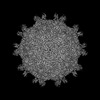 34630MC  8hbiC  8hbjC  8heeC  8hegC M: map data used to model this data C: citing same article ( |
|---|---|
| Similar structure data |


-Links
 PDBj
PDBj


- Assembly
| Deposited unit | 
|
|---|---|
| 1 | x 60
|
| 2 |
|
| 3 | x 5
|
| 4 | x 6
|
| 5 | 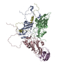
|
| Symmetry | Point symmetry: (Schoenflies symbol: I (icosahedral)) |
-Components
| #1: Protein | Mass: 23111.094 Da / Num. of mol.: 1 / Source method: isolated from a natural source / Source: (natural) Foot-and-mouth disease virus A / References: UniProt: A0A7D5BJ70 |
|---|---|
| #2: Protein | Mass: 24575.779 Da / Num. of mol.: 1 / Source method: isolated from a natural source / Source: (natural) Foot-and-mouth disease virus A / References: UniProt: A0A7D5BJ70 |
| #3: Protein | Mass: 24284.121 Da / Num. of mol.: 1 / Source method: isolated from a natural source / Source: (natural) Foot-and-mouth disease virus A / References: UniProt: A0A7D5BJ70 |
| #4: Protein | Mass: 8778.129 Da / Num. of mol.: 1 / Source method: isolated from a natural source / Source: (natural) Foot-and-mouth disease virus A / References: UniProt: A0A7D5BJ70 |
| #5: Protein | Mass: 12753.181 Da / Num. of mol.: 1 Source method: isolated from a genetically manipulated source Source: (gene. exp.) Production host: Yeast centromeric expression vector p416TEF (others) |
| Has protein modification | N |
-Experimental details
-Experiment
| Experiment | Method: ELECTRON MICROSCOPY |
|---|---|
| EM experiment | Aggregation state: PARTICLE / 3D reconstruction method: single particle reconstruction |
- Sample preparation
| Component | Name: FMDV capsid protein in complex with M678F nab / Type: COMPLEX / Entity ID: all / Source: MULTIPLE SOURCES |
|---|---|
| Source (natural) | Organism: Foot-and-mouth disease virus A |
| Buffer solution | pH: 7 |
| Specimen | Embedding applied: NO / Shadowing applied: NO / Staining applied: NO / Vitrification applied: YES |
| Vitrification | Cryogen name: ETHANE |
- Electron microscopy imaging
| Experimental equipment |  Model: Talos Arctica / Image courtesy: FEI Company |
|---|---|
| Microscopy | Model: FEI TALOS ARCTICA |
| Electron gun | Electron source:  FIELD EMISSION GUN / Accelerating voltage: 200 kV / Illumination mode: FLOOD BEAM FIELD EMISSION GUN / Accelerating voltage: 200 kV / Illumination mode: FLOOD BEAM |
| Electron lens | Mode: DIFFRACTION / Nominal defocus max: 2800 nm / Nominal defocus min: 1200 nm |
| Image recording | Electron dose: 30 e/Å2 / Film or detector model: GATAN K2 SUMMIT (4k x 4k) |
- Processing
| Software | Name: PHENIX / Version: 1.17.1_3660: / Classification: refinement | ||||||||||||||||||||||||
|---|---|---|---|---|---|---|---|---|---|---|---|---|---|---|---|---|---|---|---|---|---|---|---|---|---|
| EM software | Name: PHENIX / Category: model refinement | ||||||||||||||||||||||||
| CTF correction | Type: PHASE FLIPPING ONLY | ||||||||||||||||||||||||
| 3D reconstruction | Resolution: 3.6 Å / Resolution method: FSC 0.143 CUT-OFF / Num. of particles: 46708 / Symmetry type: POINT | ||||||||||||||||||||||||
| Refine LS restraints |
|
