 Movie
Movie Controller

[English] 日本語
 Yorodumi
- PDB-8h9h: Crystal structure of ZBTB7A in complex with GACCC-containing sequence -
+ Open data

- Basic information
| Entry | Database: PDB / ID: 8h9h | ||||||
|---|---|---|---|---|---|---|---|
| Title | Crystal structure of ZBTB7A in complex with GACCC-containing sequence | ||||||
 Components Components |
| ||||||
 Keywords Keywords | DNA BINDING PROTEIN / ZBTB7A / primed to naive transition / transcription regulation | ||||||
| Function / homology |  Function and homology information Function and homology informationregulation of transcription regulatory region DNA binding / DNA-dependent protein kinase complex / negative regulation of androgen receptor signaling pathway / double-strand break repair via classical nonhomologous end joining / regulation of DNA-binding transcription factor activity / regulation of glycolytic process / nuclear androgen receptor binding / erythrocyte maturation / regulation of alternative mRNA splicing, via spliceosome / histone acetyltransferase binding ...regulation of transcription regulatory region DNA binding / DNA-dependent protein kinase complex / negative regulation of androgen receptor signaling pathway / double-strand break repair via classical nonhomologous end joining / regulation of DNA-binding transcription factor activity / regulation of glycolytic process / nuclear androgen receptor binding / erythrocyte maturation / regulation of alternative mRNA splicing, via spliceosome / histone acetyltransferase binding / SMAD binding / fat cell differentiation / negative regulation of Notch signaling pathway / protein localization to nucleus / B cell differentiation / transcription corepressor binding / negative regulation of transforming growth factor beta receptor signaling pathway / positive regulation of NF-kappaB transcription factor activity / DNA-binding transcription repressor activity, RNA polymerase II-specific / sequence-specific double-stranded DNA binding / site of double-strand break / chromatin organization / regulation of apoptotic process / DNA-binding transcription factor binding / DNA-binding transcription factor activity, RNA polymerase II-specific / RNA polymerase II cis-regulatory region sequence-specific DNA binding / chromatin remodeling / DNA-binding transcription factor activity / negative regulation of DNA-templated transcription / DNA-templated transcription / regulation of transcription by RNA polymerase II / negative regulation of transcription by RNA polymerase II / DNA binding / zinc ion binding / nucleus / cytoplasm Similarity search - Function | ||||||
| Biological species |  Homo sapiens (human) Homo sapiens (human) | ||||||
| Method |  X-RAY DIFFRACTION / SYNCHROTRON / MOLECULAR REPLACEMENT / Resolution: 2.2 Å X-RAY DIFFRACTION / SYNCHROTRON / MOLECULAR REPLACEMENT / Resolution: 2.2 Å | ||||||
 Authors Authors | Yang, Y. | ||||||
| Funding support | 1items
| ||||||
 Citation Citation | Journal: Febs J. / Year: 2023 Title: ZBTB7A regulates primed-to-naive transition of pluripotent stem cells via recognition of the PNT-associated sequence by zinc fingers 1-2 and recognition of gamma-globin -200 gene element by zinc fingers 1-4. Authors: Yang, Y. / Xiao, L. / Xue, Y. / Idris, M.O. / Liu, J. / Pei, D. / Shi, Y. / Liao, B. / Li, F. | ||||||
| History |
|
- Structure visualization
| Structure viewer | Molecule: MolmilJmol/JSmol |
|---|
- Downloads & links
-Download
| PDBx/mmCIF format | 8h9h.cif.gz | 94.2 KB | Display | PDBx/mmCIF format |
|---|---|---|---|---|
| PDB format | pdb8h9h.ent.gz | 66.8 KB | Display | PDB format |
| PDBx/mmJSON format | 8h9h.json.gz | Tree view | PDBx/mmJSON format | |
| Others |  Other downloads Other downloads |
-Validation report
| Summary document | 8h9h_validation.pdf.gz | 2.2 MB | Display | wwPDB validaton report |
|---|---|---|---|---|
| Full document | 8h9h_full_validation.pdf.gz | 2.2 MB | Display | |
| Data in XML | 8h9h_validation.xml.gz | 7.2 KB | Display | |
| Data in CIF | 8h9h_validation.cif.gz | 9 KB | Display | |
| Arichive directory | https://data.pdbj.org/pub/pdb/validation_reports/h9/8h9hftp://data.pdbj.org/pub/pdb/validation_reports/h9/8h9h | HTTPS FTP |
-Related structure data
| Related structure data | 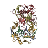 7eyiS S: Starting model for refinement |
|---|---|
| Similar structure data |
-Links
 PDBj
PDBj


- Assembly
| Deposited unit | 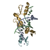
| ||||||||
|---|---|---|---|---|---|---|---|---|---|
| 1 |
| ||||||||
| Unit cell |
|
-Components
| #1: DNA chain | Mass: 3984.625 Da / Num. of mol.: 1 / Source method: obtained synthetically / Source: (synth.) | ||||||
|---|---|---|---|---|---|---|---|
| #2: DNA chain | Mass: 3957.583 Da / Num. of mol.: 1 / Source method: obtained synthetically / Source: (synth.) | ||||||
| #3: Protein | Mass: 10120.914 Da / Num. of mol.: 2 Source method: isolated from a genetically manipulated source Source: (gene. exp.) Homo sapiens (human) / Gene: ZBTB7A, FBI1, LRF, ZBTB7, ZNF857AProduction host:  References: UniProt: O95365 #4: Chemical | ChemComp-ZN /   Mass: 65.409 Da / Num. of mol.: 4 / Source method: obtained synthetically / Formula: Zn / Feature type: SUBJECT OF INVESTIGATION Mass: 65.409 Da / Num. of mol.: 4 / Source method: obtained synthetically / Formula: Zn / Feature type: SUBJECT OF INVESTIGATION#5: Water | ChemComp-HOH / |  Mass: 18.015 Da / Num. of mol.: 23 / Source method: isolated from a natural source / Formula: H2O Mass: 18.015 Da / Num. of mol.: 23 / Source method: isolated from a natural source / Formula: H2OHas ligand of interest | Y | |
-Experimental details
-Experiment
| Experiment | Method: X-RAY DIFFRACTION / Number of used crystals: 1 |
|---|
- Sample preparation
| Crystal | Density Matthews: 2.08 Å3/Da / Density % sol: 40.98 % |
|---|---|
| Crystal grow | Temperature: 293 K / Method: vapor diffusion, hanging drop / pH: 5.5 Details: 0.2 M sodium chloride, 0.1 M Bis-Tris, pH 5.5, 25% w/v PEG 3350 |
-Data collection
| Diffraction | Mean temperature: 100 K / Serial crystal experiment: N | ||||||||||||||||||||||||||||||
|---|---|---|---|---|---|---|---|---|---|---|---|---|---|---|---|---|---|---|---|---|---|---|---|---|---|---|---|---|---|---|---|
| Diffraction source | Source: SYNCHROTRON / Site: SSRF  / Beamline: BL19U1 / Wavelength: 0.9785 Å / Beamline: BL19U1 / Wavelength: 0.9785 Å | ||||||||||||||||||||||||||||||
| Detector | Type: DECTRIS PILATUS 6M / Detector: PIXEL / Date: Sep 19, 2021 | ||||||||||||||||||||||||||||||
| Radiation | Protocol: SINGLE WAVELENGTH / Monochromatic (M) / Laue (L): M / Scattering type: x-ray | ||||||||||||||||||||||||||||||
| Radiation wavelength | Wavelength: 0.9785 Å / Relative weight: 1 | ||||||||||||||||||||||||||||||
| Reflection | Resolution: 2.2→48.3 Å / Num. obs: 23116 / % possible obs: 100 % / Redundancy: 36 % / Biso Wilson estimate: 59.71 Å2 / CC1/2: 0.997 / Rmerge(I) obs: 0.131 / Rpim(I) all: 0.022 / Rrim(I) all: 0.133 / Net I/σ(I): 17.6 | ||||||||||||||||||||||||||||||
| Reflection shell | Diffraction-ID: 1
|
- Processing
| Software |
| ||||||||||||||||||||||||||||||||||||||||||||||||||||||||||||||||||||||||||||||||||||||||||||||||||||||||||||||||||||||||||||||||||||||||||||||||||||||||||||||||||||||||||||||||||||||||||||||||||||||||
|---|---|---|---|---|---|---|---|---|---|---|---|---|---|---|---|---|---|---|---|---|---|---|---|---|---|---|---|---|---|---|---|---|---|---|---|---|---|---|---|---|---|---|---|---|---|---|---|---|---|---|---|---|---|---|---|---|---|---|---|---|---|---|---|---|---|---|---|---|---|---|---|---|---|---|---|---|---|---|---|---|---|---|---|---|---|---|---|---|---|---|---|---|---|---|---|---|---|---|---|---|---|---|---|---|---|---|---|---|---|---|---|---|---|---|---|---|---|---|---|---|---|---|---|---|---|---|---|---|---|---|---|---|---|---|---|---|---|---|---|---|---|---|---|---|---|---|---|---|---|---|---|---|---|---|---|---|---|---|---|---|---|---|---|---|---|---|---|---|---|---|---|---|---|---|---|---|---|---|---|---|---|---|---|---|---|---|---|---|---|---|---|---|---|---|---|---|---|---|---|---|---|
| Refinement | Method to determine structure: MOLECULAR REPLACEMENT Starting model: 7EYI Resolution: 2.2→48.3 Å / SU ML: 0.38 / Cross valid method: THROUGHOUT / σ(F): 1.34 / Phase error: 33.54 / Stereochemistry target values: ML
| ||||||||||||||||||||||||||||||||||||||||||||||||||||||||||||||||||||||||||||||||||||||||||||||||||||||||||||||||||||||||||||||||||||||||||||||||||||||||||||||||||||||||||||||||||||||||||||||||||||||||
| Solvent computation | Shrinkage radii: 0.9 Å / VDW probe radii: 1.1 Å / Solvent model: FLAT BULK SOLVENT MODEL | ||||||||||||||||||||||||||||||||||||||||||||||||||||||||||||||||||||||||||||||||||||||||||||||||||||||||||||||||||||||||||||||||||||||||||||||||||||||||||||||||||||||||||||||||||||||||||||||||||||||||
| Displacement parameters | Biso max: 182.04 Å2 / Biso mean: 83.6559 Å2 / Biso min: 38.45 Å2 | ||||||||||||||||||||||||||||||||||||||||||||||||||||||||||||||||||||||||||||||||||||||||||||||||||||||||||||||||||||||||||||||||||||||||||||||||||||||||||||||||||||||||||||||||||||||||||||||||||||||||
| Refinement step | Cycle: final / Resolution: 2.2→48.3 Å
| ||||||||||||||||||||||||||||||||||||||||||||||||||||||||||||||||||||||||||||||||||||||||||||||||||||||||||||||||||||||||||||||||||||||||||||||||||||||||||||||||||||||||||||||||||||||||||||||||||||||||
| LS refinement shell | Refine-ID: X-RAY DIFFRACTION / Rfactor Rfree error: 0 / Total num. of bins used: 9 / % reflection obs: 100 %
| ||||||||||||||||||||||||||||||||||||||||||||||||||||||||||||||||||||||||||||||||||||||||||||||||||||||||||||||||||||||||||||||||||||||||||||||||||||||||||||||||||||||||||||||||||||||||||||||||||||||||
| Refinement TLS params. | Method: refined / Refine-ID: X-RAY DIFFRACTION
| ||||||||||||||||||||||||||||||||||||||||||||||||||||||||||||||||||||||||||||||||||||||||||||||||||||||||||||||||||||||||||||||||||||||||||||||||||||||||||||||||||||||||||||||||||||||||||||||||||||||||
| Refinement TLS group |
|
