 Movie
Movie Controller

[English] 日本語
 Yorodumi
- PDB-8h9a: Crystal structure of chemically modified E. coli ThrS catalytic d... -
+ Open data

- Basic information
| Entry | Database: PDB / ID: 8h9a | ||||||
|---|---|---|---|---|---|---|---|
| Title | Crystal structure of chemically modified E. coli ThrS catalytic domain 2 | ||||||
 Components Components | Threonine--tRNA ligase | ||||||
 Keywords Keywords | LIGASE / Threonine--tRNA ligase | ||||||
| Function / homology |  Function and homology information Function and homology informationthreonine-tRNA ligase / threonyl-tRNA aminoacylation / threonine-tRNA ligase activity / tRNA binding / ATP binding / metal ion binding / cytosol Similarity search - Function | ||||||
| Biological species |  | ||||||
| Method |  X-RAY DIFFRACTION / SYNCHROTRON / MOLECULAR REPLACEMENT / Resolution: 1.9 Å X-RAY DIFFRACTION / SYNCHROTRON / MOLECULAR REPLACEMENT / Resolution: 1.9 Å | ||||||
 Authors Authors | Qiao, H. / Xia, M. / Wang, J. / Fang, P. | ||||||
| Funding support |  China, 1items China, 1items
| ||||||
 Citation Citation | Journal: Commun Biol / Year: 2023 Title: Tyrosine-targeted covalent inhibition of a tRNA synthetase aided by zinc ion. Authors: Qiao, H. / Xia, M. / Cheng, Y. / Zhou, J. / Zheng, L. / Li, W. / Wang, J. / Fang, P. | ||||||
| History |
|
- Structure visualization
| Structure viewer | Molecule: MolmilJmol/JSmol |
|---|
- Downloads & links
-Download
| PDBx/mmCIF format | 8h9a.cif.gz | 186.2 KB | Display | PDBx/mmCIF format |
|---|---|---|---|---|
| PDB format | pdb8h9a.ent.gz | 144.2 KB | Display | PDB format |
| PDBx/mmJSON format | 8h9a.json.gz | Tree view | PDBx/mmJSON format | |
| Others |  Other downloads Other downloads |
-Validation report
| Arichive directory | https://data.pdbj.org/pub/pdb/validation_reports/h9/8h9aftp://data.pdbj.org/pub/pdb/validation_reports/h9/8h9a | HTTPS FTP |
|---|
-Related structure data
| Related structure data |  8h98C  8h99C  8h9bC  8h9cC 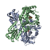 1fyfS S: Starting model for refinement C: citing same article ( |
|---|---|
| Similar structure data |
-Links
 PDBj
PDBj


- Assembly
| Deposited unit | 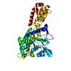
| ||||||||
|---|---|---|---|---|---|---|---|---|---|
| 1 | 
| ||||||||
| Unit cell |
|
-Components
| #1: Protein | Mass: 47893.523 Da / Num. of mol.: 1 / Fragment: UNP residues 242-642 / Mutation: Y462K Source method: isolated from a genetically manipulated source Source: (gene. exp.) |
|---|---|
| #2: Chemical | ChemComp-X5V /   Type: L-peptide NH3 amino terminus / Mass: 376.318 Da / Num. of mol.: 1 Type: L-peptide NH3 amino terminus / Mass: 376.318 Da / Num. of mol.: 1Source method: isolated from a genetically manipulated source Formula: C17H16N2O8 / Feature type: SUBJECT OF INVESTIGATION |
| #3: Chemical | ChemComp-ZN /   Mass: 65.409 Da / Num. of mol.: 1 / Source method: obtained synthetically / Formula: Zn Mass: 65.409 Da / Num. of mol.: 1 / Source method: obtained synthetically / Formula: Zn |
| #4: Water | ChemComp-HOH /  Mass: 18.015 Da / Num. of mol.: 305 / Source method: isolated from a natural source / Formula: H2O Mass: 18.015 Da / Num. of mol.: 305 / Source method: isolated from a natural source / Formula: H2O |
| Has ligand of interest | Y |
| Has protein modification | Y |
-Experimental details
-Experiment
| Experiment | Method: X-RAY DIFFRACTION / Number of used crystals: 1 |
|---|
- Sample preparation
| Crystal | Density Matthews: 3.08 Å3/Da / Density % sol: 60.12 % |
|---|---|
| Crystal grow | Temperature: 291 K / Method: vapor diffusion, sitting drop Details: 2.0 M smmonium sulfate, 0.15 M sodium citrate, pH 5.5 |
-Data collection
| Diffraction | Mean temperature: 100 K / Serial crystal experiment: N |
|---|---|
| Diffraction source | Source: SYNCHROTRON / Site: SSRF / Beamline: BL10U2 / Wavelength: 0.97857 Å |
| Detector | Type: DECTRIS EIGER X 16M / Detector: PIXEL / Date: Sep 30, 2022 |
| Radiation | Protocol: SINGLE WAVELENGTH / Monochromatic (M) / Laue (L): M / Scattering type: x-ray |
| Radiation wavelength | Wavelength: 0.97857 Å / Relative weight: 1 |
| Reflection | Resolution: 1.89→50 Å / Num. obs: 25514 / % possible obs: 98.1 % / Redundancy: 8.5 % / Rmerge(I) obs: 0.096 / Net I/σ(I): 10.1 |
| Reflection shell | Resolution: 1.89→1.94 Å / Rmerge(I) obs: 0.606 / Num. unique obs: 2980 |
- Processing
| Software |
| ||||||||||||||||||||||||||||||||||||||||||||||||||||||||||||||||||||||
|---|---|---|---|---|---|---|---|---|---|---|---|---|---|---|---|---|---|---|---|---|---|---|---|---|---|---|---|---|---|---|---|---|---|---|---|---|---|---|---|---|---|---|---|---|---|---|---|---|---|---|---|---|---|---|---|---|---|---|---|---|---|---|---|---|---|---|---|---|---|---|---|
| Refinement | Method to determine structure: MOLECULAR REPLACEMENT Starting model: PDB entry 1FYF Resolution: 1.9→42.87 Å / SU ML: 0.16 / Cross valid method: THROUGHOUT / σ(F): 1.36 / Phase error: 25.67 / Stereochemistry target values: ML
| ||||||||||||||||||||||||||||||||||||||||||||||||||||||||||||||||||||||
| Solvent computation | Shrinkage radii: 0.9 Å / VDW probe radii: 1.1 Å / Solvent model: FLAT BULK SOLVENT MODEL | ||||||||||||||||||||||||||||||||||||||||||||||||||||||||||||||||||||||
| Displacement parameters | Biso max: 141.36 Å2 / Biso mean: 28.9811 Å2 / Biso min: 7.37 Å2 | ||||||||||||||||||||||||||||||||||||||||||||||||||||||||||||||||||||||
| Refinement step | Cycle: final / Resolution: 1.9→42.87 Å
| ||||||||||||||||||||||||||||||||||||||||||||||||||||||||||||||||||||||
| LS refinement shell | Refine-ID: X-RAY DIFFRACTION / Rfactor Rfree error: 0 / Total num. of bins used: 9
|
