 Movie
Movie Controller

[English] 日本語
 Yorodumi
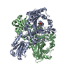
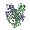
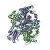
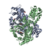
- PDB-1fyf: CRYSTAL STRUCTURE OF A TRUNCATED FORM OF THREONYL-TRNA SYNTHETASE... -
+ Open data

- Basic information
| Entry | Database: PDB / ID: 1fyf | ||||||
|---|---|---|---|---|---|---|---|
| Title | CRYSTAL STRUCTURE OF A TRUNCATED FORM OF THREONYL-TRNA SYNTHETASE COMPLEXED WITH A SERYL ADENYLATE ANALOG | ||||||
 Components Components | THREONYL-TRNA SYNTHETASE | ||||||
 Keywords Keywords | LIGASE / AMINO ACID RECOGNITION / ZINC ION / TRNA-SYNTHETASE / ADENYLATE ANALOG / DELETION MUTANT | ||||||
| Function / homology |  Function and homology information Function and homology informationaminoacyl-tRNA ligase activity / tRNA aminoacylation / threonine-tRNA ligase / threonyl-tRNA aminoacylation / threonine-tRNA ligase activity / tRNA aminoacylation for protein translation / aminoacyl-tRNA deacylase activity / negative regulation of translational initiation / mRNA regulatory element binding translation repressor activity / mRNA 5'-UTR binding ...aminoacyl-tRNA ligase activity / tRNA aminoacylation / threonine-tRNA ligase / threonyl-tRNA aminoacylation / threonine-tRNA ligase activity / tRNA aminoacylation for protein translation / aminoacyl-tRNA deacylase activity / negative regulation of translational initiation / mRNA regulatory element binding translation repressor activity / mRNA 5'-UTR binding / regulation of translation / tRNA binding / response to antibiotic / protein homodimerization activity / RNA binding / zinc ion binding / ATP binding / cytosol / cytoplasm Similarity search - Function | ||||||
| Biological species |  | ||||||
| Method |  X-RAY DIFFRACTION / SYNCHROTRON / Resolution: 1.65 Å X-RAY DIFFRACTION / SYNCHROTRON / Resolution: 1.65 Å | ||||||
 Authors Authors | Sankaranarayanan, R. / Dock-Bregeon, A.C. / Moras, D. | ||||||
 Citation Citation | Journal: Cell(Cambridge,Mass.) / Year: 2000 Title: Transfer RNA-mediated editing in threonyl-tRNA synthetase. The class II solution to the double discrimination problem. Authors: Dock-Bregeon, A. / Sankaranarayanan, R. / Romby, P. / Caillet, J. / Springer, M. / Rees, B. / Francklyn, C.S. / Ehresmann, C. / Moras, D. #1: Journal: Cell(Cambridge,Mass.) / Year: 1999Title: The Structure of Threonyl-tRNA Synthetase-tRNA(Thr) Complex Enlightens its Repressor Activity and Reveals an Essential Zinc Ion in the Active Site Authors: Sankaranarayanan, R. / Dock-Bregeon, A.C. / Romby, P. / Caillet, J. / Springer, M. / Rees, B. / Ehresmann, C. / Ehresmann, B. / Moras, D. #2: Journal: Nat.Struct.Biol. / Year: 2000Title: Zinc Ion Mediated Amino Acid Discrimination by Threonyl-tRNA Synthetase Authors: Sankaranarayanan, R. / Dock-Bregeon, A.C. / Rees, B. / Bovee, M. / Caillet, J. / Romby, P. / Francklyn, C.S. / Moras, D. | ||||||
| History |
|
- Structure visualization
| Structure viewer | Molecule: MolmilJmol/JSmol |
|---|
- Downloads & links
-Download
| PDBx/mmCIF format | 1fyf.cif.gz | 185.7 KB | Display | PDBx/mmCIF format |
|---|---|---|---|---|
| PDB format | pdb1fyf.ent.gz | 144.2 KB | Display | PDB format |
| PDBx/mmJSON format | 1fyf.json.gz | Tree view | PDBx/mmJSON format | |
| Others |  Other downloads Other downloads |
-Validation report
| Arichive directory | https://data.pdbj.org/pub/pdb/validation_reports/fy/1fyfftp://data.pdbj.org/pub/pdb/validation_reports/fy/1fyf | HTTPS FTP |
|---|
-Related structure data
| Related structure data | |
|---|---|
| Similar structure data |


-Links
 PDBj
PDBj


- Assembly
| Deposited unit | 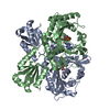
| ||||||||
|---|---|---|---|---|---|---|---|---|---|
| 1 |
| ||||||||
| Unit cell |
| ||||||||
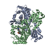
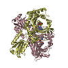
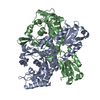
| Details | The biological assembly is a dimer which is in the asymmetric unit. |
-Components
| #1: Protein | Mass: 46725.180 Da / Num. of mol.: 2 Fragment: CATALYTIC AND ANTICODON BINDING DOMAINS (RESIDUES 242-642) Source method: isolated from a genetically manipulated source Source: (gene. exp.) #2: Chemical |   Mass: 65.409 Da / Num. of mol.: 2 / Source method: obtained synthetically / Formula: Zn Mass: 65.409 Da / Num. of mol.: 2 / Source method: obtained synthetically / Formula: Zn#3: Chemical | 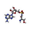  Mass: 433.397 Da / Num. of mol.: 2 / Source method: obtained synthetically / Formula: C13H19N7O8S Mass: 433.397 Da / Num. of mol.: 2 / Source method: obtained synthetically / Formula: C13H19N7O8S#4: Water | ChemComp-HOH / |  Mass: 18.015 Da / Num. of mol.: 534 / Source method: isolated from a natural source / Formula: H2O Mass: 18.015 Da / Num. of mol.: 534 / Source method: isolated from a natural source / Formula: H2O |
|---|
-Experimental details
-Experiment
| Experiment | Method: X-RAY DIFFRACTION / Number of used crystals: 1 |
|---|
- Sample preparation
| Crystal | Density Matthews: 2.94 Å3/Da / Density % sol: 58.14 % | ||||||||||||||||||||
|---|---|---|---|---|---|---|---|---|---|---|---|---|---|---|---|---|---|---|---|---|---|
| Crystal grow | Temperature: 277 K / Method: vapor diffusion / pH: 6.5 Details: PEG 4000, ammonium acetate, magnesium chloride, pH 6.5, VAPOR DIFFUSION, temperature 277.0K | ||||||||||||||||||||
| Crystal grow | *PLUS Method: vapor diffusion, hanging drop | ||||||||||||||||||||
| Components of the solutions | *PLUS
|
-Data collection
| Diffraction | Mean temperature: 120 K |
|---|---|
| Diffraction source | Source: SYNCHROTRON / Site: EMBL/DESY, HAMBURG  / Beamline: BW7B / Wavelength: 0.8439 / Beamline: BW7B / Wavelength: 0.8439 |
| Detector | Type: MARRESEARCH / Detector: IMAGE PLATE / Date: Jun 18, 1999 |
| Radiation | Protocol: SINGLE WAVELENGTH / Monochromatic (M) / Laue (L): M / Scattering type: x-ray |
| Radiation wavelength | Wavelength: 0.8439 Å / Relative weight: 1 |
| Reflection | Resolution: 1.65→20 Å / Num. all: 127385 / Num. obs: 127385 / % possible obs: 95.6 % / Redundancy: 3.3 % / Biso Wilson estimate: 18.7 Å2 / Rmerge(I) obs: 0.053 / Net I/σ(I): 21.7 |
| Reflection shell | Resolution: 1.65→1.69 Å / Redundancy: 2.7 % / Rmerge(I) obs: 0.276 / Num. unique all: 7268 / % possible all: 82.9 |
| Reflection | *PLUS Num. measured all: 431526 |
| Reflection shell | *PLUS % possible obs: 82.9 % |
- Processing
| Software |
| ||||||||||||||||||||||||||||||||||||
|---|---|---|---|---|---|---|---|---|---|---|---|---|---|---|---|---|---|---|---|---|---|---|---|---|---|---|---|---|---|---|---|---|---|---|---|---|---|
| Refinement | Resolution: 1.65→19.93 Å / Rfactor Rfree error: 0.003 / Data cutoff high absF: 2345367.94 / Data cutoff low absF: 0 / Isotropic thermal model: RESTRAINED / Cross valid method: THROUGHOUT / σ(F): 2 / Stereochemistry target values: Engh & Huber
| ||||||||||||||||||||||||||||||||||||
| Solvent computation | Solvent model: FLAT MODEL / Bsol: 44.55 Å2 / ksol: 0.381 e/Å3 | ||||||||||||||||||||||||||||||||||||
| Displacement parameters | Biso mean: 22.8 Å2
| ||||||||||||||||||||||||||||||||||||
| Refine analyze |
| ||||||||||||||||||||||||||||||||||||
| Refinement step | Cycle: LAST / Resolution: 1.65→19.93 Å
| ||||||||||||||||||||||||||||||||||||
| Refine LS restraints |
| ||||||||||||||||||||||||||||||||||||
| LS refinement shell | Resolution: 1.65→1.75 Å / Rfactor Rfree error: 0.009 / Total num. of bins used: 6
| ||||||||||||||||||||||||||||||||||||
| Xplor file |
| ||||||||||||||||||||||||||||||||||||
| Software | *PLUS Name: CNS / Version: 1 / Classification: refinement | ||||||||||||||||||||||||||||||||||||
| Refine LS restraints | *PLUS
|
