 Movie
Movie Controller


+ Open data

- Basic information
| Entry | Database: PDB / ID: 8h8j | ||||||
|---|---|---|---|---|---|---|---|
| Title | Lodoxamide-bound GPR35 in complex with G13 | ||||||
 Components Components |
| ||||||
 Keywords Keywords | MEMBRANE PROTEIN / GPR35 / G13 / Lodoxamide | ||||||
| Function / homology |  Function and homology information Function and homology informationD5 dopamine receptor binding / negative regulation of voltage-gated calcium channel activity / leukocyte adhesion to vascular endothelial cell / regulation of fibroblast migration / Rho-activating G protein-coupled receptor signaling pathway / chemokine receptor activity / C-X-C chemokine receptor activity / Class A/1 (Rhodopsin-like receptors) / regulation of small GTPase mediated signal transduction / NRAGE signals death through JNK ...D5 dopamine receptor binding / negative regulation of voltage-gated calcium channel activity / leukocyte adhesion to vascular endothelial cell / regulation of fibroblast migration / Rho-activating G protein-coupled receptor signaling pathway / chemokine receptor activity / C-X-C chemokine receptor activity / Class A/1 (Rhodopsin-like receptors) / regulation of small GTPase mediated signal transduction / NRAGE signals death through JNK / branching involved in blood vessel morphogenesis / CDC42 GTPase cycle / negative regulation of cardiac muscle cell apoptotic process / monocyte chemotaxis / regulation of postsynapse assembly / Rho protein signal transduction / neutrophil chemotaxis / RAC1 GTPase cycle / cytoskeleton organization / guanyl-nucleotide exchange factor activity / response to ischemia / chemokine-mediated signaling pathway / brush border membrane / platelet activation / regulation of blood pressure / G protein-coupled receptor activity / G-protein beta/gamma-subunit complex binding / adenylate cyclase-modulating G protein-coupled receptor signaling pathway / adenylate cyclase-inhibiting G protein-coupled receptor signaling pathway / Olfactory Signaling Pathway / Activation of the phototransduction cascade / G protein-coupled acetylcholine receptor signaling pathway / G beta:gamma signalling through PLC beta / Presynaptic function of Kainate receptors / Thromboxane signalling through TP receptor / Activation of G protein gated Potassium channels / Inhibition of voltage gated Ca2+ channels via Gbeta/gamma subunits / G-protein activation / Glucagon signaling in metabolic regulation / Prostacyclin signalling through prostacyclin receptor / G beta:gamma signalling through CDC42 / Synthesis, secretion, and inactivation of Glucagon-like Peptide-1 (GLP-1) / melanosome / G beta:gamma signalling through BTK / photoreceptor disc membrane / ADP signalling through P2Y purinoceptor 12 / Glucagon-type ligand receptors / Sensory perception of sweet, bitter, and umami (glutamate) taste / Adrenaline,noradrenaline inhibits insulin secretion / Vasopressin regulates renal water homeostasis via Aquaporins / Glucagon-like Peptide-1 (GLP1) regulates insulin secretion / G alpha (z) signalling events / cellular response to catecholamine stimulus / ADP signalling through P2Y purinoceptor 1 / regulation of cell shape / ADORA2B mediated anti-inflammatory cytokines production / G beta:gamma signalling through PI3Kgamma / adenylate cyclase-activating dopamine receptor signaling pathway / Cooperation of PDCL (PhLP1) and TRiC/CCT in G-protein beta folding / GPER1 signaling / cellular response to prostaglandin E stimulus / heterotrimeric G-protein complex / G alpha (12/13) signalling events / G-protein beta-subunit binding / Inactivation, recovery and regulation of the phototransduction cascade / extracellular vesicle / sensory perception of taste / Thrombin signalling through proteinase activated receptors (PARs) / adenylate cyclase-activating G protein-coupled receptor signaling pathway / signaling receptor complex adaptor activity / positive regulation of cytosolic calcium ion concentration / retina development in camera-type eye / GTPase binding / fibroblast proliferation / G protein activity / Ca2+ pathway / High laminar flow shear stress activates signaling by PIEZO1 and PECAM1:CDH5:KDR in endothelial cells / G alpha (i) signalling events / G alpha (s) signalling events / phospholipase C-activating G protein-coupled receptor signaling pathway / G alpha (q) signalling events / in utero embryonic development / Ras protein signal transduction / cell differentiation / Extra-nuclear estrogen signaling / cell population proliferation / postsynapse / G protein-coupled receptor signaling pathway / lysosomal membrane / focal adhesion / GTPase activity / synapse / GTP binding / protein-containing complex binding / signal transduction / extracellular exosome / membrane / metal ion binding / nucleus / plasma membrane Similarity search - Function | ||||||
| Biological species |  Homo sapiens (human) Homo sapiens (human) | ||||||
| Method | ELECTRON MICROSCOPY / single particle reconstruction / cryo EM / Resolution: 3.2 Å | ||||||
 Authors Authors | Yuan, Q. / Duan, J. / Liu, Q. / Xu, H.E. / Jiang, Y. | ||||||
| Funding support |  China, 1items China, 1items
| ||||||
 Citation Citation | Journal: Cell Discov / Year: 2022 Title: Insights into divalent cation regulation and G-coupling of orphan receptor GPR35. Authors: Jia Duan / Qiufeng Liu / Qingning Yuan / Yujie Ji / Shengnan Zhu / Yangxia Tan / Xinheng He / Youwei Xu / Jingjing Shi / Xi Cheng / Hualiang Jiang / H Eric Xu / Yi Jiang / Abstract: Endogenous ions play important roles in the function and pharmacology of G protein-coupled receptors (GPCRs) with limited atomic evidence. In addition, compared with G protein subtypes G, G, and G, ...Endogenous ions play important roles in the function and pharmacology of G protein-coupled receptors (GPCRs) with limited atomic evidence. In addition, compared with G protein subtypes G, G, and G, insufficient structural evidence is accessible to understand the coupling mechanism of G protein by GPCRs. Orphan receptor GPR35, which is predominantly expressed in the gastrointestinal tract and is closely related to inflammatory bowel diseases (IBDs), stands out as a prototypical receptor for investigating ionic modulation and G coupling. Here we report a cryo-electron microscopy structure of G-coupled GPR35 bound to an anti-allergic drug, lodoxamide. This structure reveals a novel divalent cation coordination site and a unique ionic regulatory mode of GPR35 and also presents a highly positively charged binding pocket and the complementary electrostatic ligand recognition mode, which explain the promiscuity of acidic ligand binding by GPR35. Structural comparison of the GPR35-G complex with other G protein subtypes-coupled GPCRs reveals a notable movement of the C-terminus of α5 helix of the Gα subunit towards the receptor core and the least outward displacement of the cytoplasmic end of GPR35 TM6. A featured 'methionine pocket' contributes to the G coupling by GPR35. Together, our findings provide a structural basis for divalent cation modulation, ligand recognition, and subsequent G protein coupling of GPR35 and offer a new opportunity for designing GPR35-targeted drugs for the treatment of IBDs. | ||||||
| History |
|
- Structure visualization
| Structure viewer | Molecule: MolmilJmol/JSmol |
|---|
- Downloads & links
-Download
| PDBx/mmCIF format | 8h8j.cif.gz | 216.7 KB | Display | PDBx/mmCIF format |
|---|---|---|---|---|
| PDB format | pdb8h8j.ent.gz | 164.9 KB | Display | PDB format |
| PDBx/mmJSON format | 8h8j.json.gz | Tree view | PDBx/mmJSON format | |
| Others |  Other downloads Other downloads |
-Validation report
| Arichive directory | https://data.pdbj.org/pub/pdb/validation_reports/h8/8h8jftp://data.pdbj.org/pub/pdb/validation_reports/h8/8h8j | HTTPS FTP |
|---|
-Related structure data
| Related structure data |  34549MC M: map data used to model this data C: citing same article ( |
|---|---|
| Similar structure data |
-Links
 PDBj
PDBj


- Assembly
| Deposited unit | 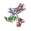
|
|---|---|
| 1 |
|
-Components
-Guanine nucleotide-binding protein ... , 3 types, 3 molecules ABG
| #1: Protein | Mass: 42423.633 Da / Num. of mol.: 1 Source method: isolated from a genetically manipulated source Source: (gene. exp.) Homo sapiens (human) / Gene: GNA13 / Production host:   Spodoptera frugiperda (fall armyworm) / References: UniProt: Q14344 Spodoptera frugiperda (fall armyworm) / References: UniProt: Q14344 |
|---|---|
| #2: Protein | Mass: 40226.992 Da / Num. of mol.: 1 Source method: isolated from a genetically manipulated source Source: (gene. exp.) Homo sapiens (human) / Gene: GNB1 / Production host: Spodoptera frugiperda (fall armyworm) / References: UniProt: P62873 |
| #4: Protein | Mass: 7861.143 Da / Num. of mol.: 1 Source method: isolated from a genetically manipulated source Source: (gene. exp.) Homo sapiens (human) / Gene: GNG2 / Production host: Spodoptera frugiperda (fall armyworm) / References: UniProt: P59768 |
-Protein / Antibody , 2 types, 2 molecules CH
| #3: Protein | Mass: 33972.047 Da / Num. of mol.: 1 Source method: isolated from a genetically manipulated source Source: (gene. exp.) Homo sapiens (human) / Gene: GPR35 / Production host: Spodoptera frugiperda (fall armyworm) / References: UniProt: Q9HC97 |
|---|---|
| #5: Antibody | Mass: 26277.299 Da / Num. of mol.: 1 Source method: isolated from a genetically manipulated source Source: (gene. exp.) Spodoptera frugiperda (fall armyworm) |
-Non-polymers , 3 types, 5 molecules 


| #6: Chemical | ChemComp-WYB /  Mass: 311.635 Da / Num. of mol.: 1 / Source method: obtained synthetically / Formula: C11H6ClN3O6 Mass: 311.635 Da / Num. of mol.: 1 / Source method: obtained synthetically / Formula: C11H6ClN3O6 |
|---|---|
| #7: Chemical | ChemComp-CA /  Mass: 40.078 Da / Num. of mol.: 1 / Source method: obtained synthetically / Formula: Ca Mass: 40.078 Da / Num. of mol.: 1 / Source method: obtained synthetically / Formula: Ca |
| #8: Chemical |  Mass: 386.654 Da / Num. of mol.: 3 / Source method: obtained synthetically / Formula: C27H46O Mass: 386.654 Da / Num. of mol.: 3 / Source method: obtained synthetically / Formula: C27H46O |
-Details
| Has ligand of interest | N |
|---|---|
| Has protein modification | Y |
-Experimental details
-Experiment
| Experiment | Method: ELECTRON MICROSCOPY |
|---|---|
| EM experiment | Aggregation state: PARTICLE / 3D reconstruction method: single particle reconstruction |
- Sample preparation
| Component | Name: Lodoxamide-GPR35-G13 complex / Type: COMPLEX / Entity ID: #1-#5 / Source: RECOMBINANT |
|---|---|
| Source (natural) | Organism: Homo sapiens (human) |
| Source (recombinant) | Organism: Spodoptera frugiperda (fall armyworm) |
| Buffer solution | pH: 7.04 |
| Specimen | Embedding applied: NO / Shadowing applied: NO / Staining applied: NO / Vitrification applied: YES |
| Vitrification | Cryogen name: ETHANE |
- Electron microscopy imaging
| Experimental equipment |  Model: Titan Krios / Image courtesy: FEI Company |
|---|---|
| Microscopy | Model: FEI TITAN KRIOS |
| Electron gun | Electron source:  FIELD EMISSION GUN / Accelerating voltage: 300 kV / Illumination mode: FLOOD BEAM FIELD EMISSION GUN / Accelerating voltage: 300 kV / Illumination mode: FLOOD BEAM |
| Electron lens | Mode: BRIGHT FIELD / Nominal defocus max: 2000 nm / Nominal defocus min: 800 nm |
| Image recording | Electron dose: 50 e/Å2 / Film or detector model: GATAN K3 BIOQUANTUM (6k x 4k) |
- Processing
| CTF correction | Type: PHASE FLIPPING AND AMPLITUDE CORRECTION |
|---|---|
| 3D reconstruction | Resolution: 3.2 Å / Resolution method: FSC 0.143 CUT-OFF / Num. of particles: 591246 / Symmetry type: POINT |
