National Natural Science Foundation of China (NSFC)
China
Citation
Journal: Cell Discov / Year: 2022 Title: Insights into divalent cation regulation and G-coupling of orphan receptor GPR35. Authors: Jia Duan / Qiufeng Liu / Qingning Yuan / Yujie Ji / Shengnan Zhu / Yangxia Tan / Xinheng He / Youwei Xu / Jingjing Shi / Xi Cheng / Hualiang Jiang / H Eric Xu / Yi Jiang / Abstract: Endogenous ions play important roles in the function and pharmacology of G protein-coupled receptors (GPCRs) with limited atomic evidence. In addition, compared with G protein subtypes G, G, and G, ...Endogenous ions play important roles in the function and pharmacology of G protein-coupled receptors (GPCRs) with limited atomic evidence. In addition, compared with G protein subtypes G, G, and G, insufficient structural evidence is accessible to understand the coupling mechanism of G protein by GPCRs. Orphan receptor GPR35, which is predominantly expressed in the gastrointestinal tract and is closely related to inflammatory bowel diseases (IBDs), stands out as a prototypical receptor for investigating ionic modulation and G coupling. Here we report a cryo-electron microscopy structure of G-coupled GPR35 bound to an anti-allergic drug, lodoxamide. This structure reveals a novel divalent cation coordination site and a unique ionic regulatory mode of GPR35 and also presents a highly positively charged binding pocket and the complementary electrostatic ligand recognition mode, which explain the promiscuity of acidic ligand binding by GPR35. Structural comparison of the GPR35-G complex with other G protein subtypes-coupled GPCRs reveals a notable movement of the C-terminus of α5 helix of the Gα subunit towards the receptor core and the least outward displacement of the cytoplasmic end of GPR35 TM6. A featured 'methionine pocket' contributes to the G coupling by GPR35. Together, our findings provide a structural basis for divalent cation modulation, ligand recognition, and subsequent G protein coupling of GPR35 and offer a new opportunity for designing GPR35-targeted drugs for the treatment of IBDs.
In the structure databanks used in Yorodumi, some data are registered as the other names, "COVID-19 virus" and "2019-nCoV". Here are the details of the virus and the list of structure data.
Jan 31, 2019. EMDB accession codes are about to change! (news from PDBe EMDB page)
EMDB accession codes are about to change! (news from PDBe EMDB page)
The allocation of 4 digits for EMDB accession codes will soon come to an end. Whilst these codes will remain in use, new EMDB accession codes will include an additional digit and will expand incrementally as the available range of codes is exhausted. The current 4-digit format prefixed with “EMD-” (i.e. EMD-XXXX) will advance to a 5-digit format (i.e. EMD-XXXXX), and so on. It is currently estimated that the 4-digit codes will be depleted around Spring 2019, at which point the 5-digit format will come into force.
The EM Navigator/Yorodumi systems omit the EMD- prefix.
Related info.:Q: What is EMD? / ID/Accession-code notation in Yorodumi/EM Navigator
Yorodumi is a browser for structure data from EMDB, PDB, SASBDB, etc.
This page is also the successor to EM Navigator detail page, and also detail information page/front-end page for Omokage search.
The word "yorodu" (or yorozu) is an old Japanese word meaning "ten thousand". "mi" (miru) is to see.
Related info.:EMDB / PDB / SASBDB / Comparison of 3 databanks / Yorodumi Search / Aug 31, 2016. New EM Navigator & Yorodumi / Yorodumi Papers / Jmol/JSmol / Function and homology information / Changes in new EM Navigator and Yorodumi
 Movie
Movie Controller


 Open data
 Basic information
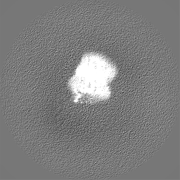
 Map data
 Sample
 Keywords
 Function and homology information
 Homo sapiens (human) /
 Authors
 China, 1 items
  Citation
 Structure visualization
 Downloads & links
 emd_34549.png
 http://ftp.pdbj.org/pub/emdb/structures/EMD-34549


 Z (Sec.)
 Y (Row.)
 X (Col.)


 Sample components
 Spodoptera frugiperda (fall armyworm)

 Processing
 Electron microscopy
 FIELD EMISSION GUN
