 Movie
Movie Controller

[English] 日本語
 Yorodumi
- PDB-8h8c: Type VI secretion system effector RhsP in its post-autoproteolysi... -
+ Open data

- Basic information
| Entry | Database: PDB / ID: 8h8c | |||||||||||||||||||||||||||||||||||||||
|---|---|---|---|---|---|---|---|---|---|---|---|---|---|---|---|---|---|---|---|---|---|---|---|---|---|---|---|---|---|---|---|---|---|---|---|---|---|---|---|---|
| Title | Type VI secretion system effector RhsP in its post-autoproteolysis and dimeric form | |||||||||||||||||||||||||||||||||||||||
 Components Components |
| |||||||||||||||||||||||||||||||||||||||
 Keywords Keywords | TOXIN / T6SS / Rhs proteins / polymorphic toxins | |||||||||||||||||||||||||||||||||||||||
| Function / homology |  Function and homology information Function and homology informationWHH domain-containing protein / A nuclease of the HNH/ENDO VII superfamily with conserved WHH / Domain of unknown function DUF6531 / Domain of unknown function (DUF6531) / : / Teneurin YD-shell / YD repeat / : / Rhs repeat-associated core Similarity search - Domain/homology | |||||||||||||||||||||||||||||||||||||||
| Biological species | Vibrio parahaemolyticus serotype O3:K6 | |||||||||||||||||||||||||||||||||||||||
| Method | ELECTRON MICROSCOPY / single particle reconstruction / cryo EM / Resolution: 3.36 Å | |||||||||||||||||||||||||||||||||||||||
 Authors Authors | Tang, L. / Dong, S.Q. / Rasheed, N. / Wu, H.W. / Zhou, N.K. / Li, H.D. / Wang, M.L. / Zheng, J. / He, J. / Chao, W.C.H. | |||||||||||||||||||||||||||||||||||||||
| Funding support |  China, 2items China, 2items
| |||||||||||||||||||||||||||||||||||||||
 Citation Citation | Journal: Cell Rep / Year: 2022 Title: Vibrio parahaemolyticus prey targeting requires autoproteolysis-triggered dimerization of the type VI secretion system effector RhsP. Authors: Le Tang / Shuqi Dong / Nadia Rasheed / Hao Weng Wu / Ningkun Zhou / Huadong Li / Meilin Wang / Jun Zheng / Jun He / William Chong Hang Chao / Abstract: The rearrangement hotspot (Rhs) repeat is an ancient giant protein fold found in all domains of life. Rhs proteins are polymorphic toxins that could either be deployed as an ABC complex or via a type ...The rearrangement hotspot (Rhs) repeat is an ancient giant protein fold found in all domains of life. Rhs proteins are polymorphic toxins that could either be deployed as an ABC complex or via a type VI secretion system (T6SS) in interbacterial competitions. To explore the mechanism of T6SS-delivered Rhs toxins, we used the gastroenteritis-associated Vibrio parahaemolyticus as a model organism and identified an Rhs toxin-immunity pair, RhsP-RhsP. Our data show that RhsP-dependent prey targeting by V. parahaemolyticus requires T6SS2. RhsP can bind to VgrG2 independently without a chaperone and spontaneously self-cleaves into three fragments. The toxic C-terminal fragment (RhsP) can bind to VgrG2 via a VgrG2-interacting region (VIR). Our electron microscopy (EM) analysis reveals that the VIR is encapsulated inside the Rhs β barrel structure and that autoproteolysis triggers a dramatic conformational change of the VIR. This alternative VIR conformation promotes RhsP dimerization, which significantly contributes to T6SS2-mediated prey targeting by V. parahaemolyticus. | |||||||||||||||||||||||||||||||||||||||
| History |
|
- Structure visualization
| Structure viewer | Molecule: MolmilJmol/JSmol |
|---|
- Downloads & links
-Download
| PDBx/mmCIF format | 8h8c.cif.gz | 396.7 KB | Display | PDBx/mmCIF format |
|---|---|---|---|---|
| PDB format | pdb8h8c.ent.gz | 307.2 KB | Display | PDB format |
| PDBx/mmJSON format | 8h8c.json.gz | Tree view | PDBx/mmJSON format | |
| Others |  Other downloads Other downloads |
-Validation report
| Arichive directory | https://data.pdbj.org/pub/pdb/validation_reports/h8/8h8cftp://data.pdbj.org/pub/pdb/validation_reports/h8/8h8c | HTTPS FTP |
|---|
-Related structure data
| Related structure data |  34542MC  8h8aC 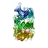 8h8bC M: map data used to model this data C: citing same article ( |
|---|---|
| Similar structure data |
-Links
 PDBj
PDBj- Assembly
| Deposited unit | 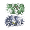
|
|---|---|
| 1 |
|
-Components
| #1: Protein | Mass: 131985.109 Da / Num. of mol.: 2 Source method: isolated from a genetically manipulated source Details: In cleaved RhsP, the cleavage at F1131/L1132 allows the VIR to re-position itself in a U-shape manner with both the N and C termini located at the lid region. Source: (gene. exp.)  Vibrio parahaemolyticus serotype O3:K6 (strain RIMD 2210633) (bacteria) Vibrio parahaemolyticus serotype O3:K6 (strain RIMD 2210633) (bacteria)Gene: VP1517 / Production host: #2: Protein | Mass: 28143.795 Da / Num. of mol.: 2 / Mutation: H1354A Source method: isolated from a genetically manipulated source Details: Cleaved RhsP in its dimeric form Source: (gene. exp.) Vibrio parahaemolyticus serotype O3:K6 (strain RIMD 2210633) (bacteria)Gene: VP1517 / Production host: Has protein modification | N | |
|---|
-Experimental details
-Experiment
| Experiment | Method: ELECTRON MICROSCOPY |
|---|---|
| EM experiment | Aggregation state: 3D ARRAY / 3D reconstruction method: single particle reconstruction |
- Sample preparation
| Component | Name: Type VI secretion system effector RhsP in its post-autoproteolysis form Type: COMPLEX / Entity ID: all / Source: MULTIPLE SOURCES |
|---|---|
| Source (natural) | Organism: Vibrio parahaemolyticus serotype O3:K6 (strain RIMD 2210633) (bacteria) |
| Source (recombinant) | Organism: |
| Buffer solution | pH: 7.5 |
| Specimen | Embedding applied: NO / Shadowing applied: NO / Staining applied: NO / Vitrification applied: YES |
| Vitrification | Cryogen name: ETHANE |
- Electron microscopy imaging
| Experimental equipment |  Model: Talos Arctica / Image courtesy: FEI Company |
|---|---|
| Microscopy | Model: FEI TALOS ARCTICA |
| Electron gun | Electron source:  FIELD EMISSION GUN / Accelerating voltage: 200 kV / Illumination mode: FLOOD BEAM FIELD EMISSION GUN / Accelerating voltage: 200 kV / Illumination mode: FLOOD BEAM |
| Electron lens | Mode: BRIGHT FIELD / Nominal defocus max: 2000 nm / Nominal defocus min: 1000 nm |
| Image recording | Electron dose: 60 e/Å2 / Film or detector model: GATAN K3 BIOQUANTUM (6k x 4k) |
- Processing
| Software | Name: PHENIX / Version: 1.17.1_3660: / Classification: refinement | ||||||||||||||||||||||||
|---|---|---|---|---|---|---|---|---|---|---|---|---|---|---|---|---|---|---|---|---|---|---|---|---|---|
| EM software | Name: PHENIX / Category: model refinement | ||||||||||||||||||||||||
| CTF correction | Type: PHASE FLIPPING AND AMPLITUDE CORRECTION | ||||||||||||||||||||||||
| 3D reconstruction | Resolution: 3.36 Å / Resolution method: FSC 0.143 CUT-OFF / Num. of particles: 212763 / Symmetry type: POINT | ||||||||||||||||||||||||
| Refine LS restraints |
|
