+ Open data
- Basic information
| Entry | Database: PDB / ID: 8h3d | ||||||
|---|---|---|---|---|---|---|---|
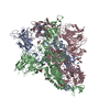
| Title | Structure of apo SARS-CoV-2 spike protein with one RBD up | ||||||
 Components Components | Spike glycoprotein,Fibritin | ||||||
 Keywords Keywords | VIRAL PROTEIN / SARS-CoV-2 / spike protein / apo | ||||||
| Function / homology |  Function and homology information Function and homology informationvirion component / symbiont-mediated disruption of host tissue / Maturation of spike protein / Translation of Structural Proteins / Virion Assembly and Release / host cell surface / host extracellular space / symbiont-mediated-mediated suppression of host tetherin activity / Induction of Cell-Cell Fusion / structural constituent of virion ...virion component / symbiont-mediated disruption of host tissue / Maturation of spike protein / Translation of Structural Proteins / Virion Assembly and Release / host cell surface / host extracellular space / symbiont-mediated-mediated suppression of host tetherin activity / Induction of Cell-Cell Fusion / structural constituent of virion / membrane fusion / entry receptor-mediated virion attachment to host cell / Attachment and Entry / host cell endoplasmic reticulum-Golgi intermediate compartment membrane / positive regulation of viral entry into host cell / receptor-mediated virion attachment to host cell / host cell surface receptor binding / symbiont-mediated suppression of host innate immune response / endocytosis involved in viral entry into host cell / receptor ligand activity / fusion of virus membrane with host plasma membrane / fusion of virus membrane with host endosome membrane / viral envelope / symbiont entry into host cell / virion attachment to host cell / SARS-CoV-2 activates/modulates innate and adaptive immune responses / host cell plasma membrane / virion membrane / identical protein binding / membrane / plasma membrane Similarity search - Function | ||||||
| Biological species |  | ||||||
| Method | ELECTRON MICROSCOPY / single particle reconstruction / cryo EM / Resolution: 3.27 Å | ||||||
 Authors Authors | Meng, F. / Wang, Q. / Xie, Y. / Ni, X. / Huang, N. | ||||||
| Funding support |  China, 1items China, 1items
| ||||||
 Citation Citation |  Journal: ACS Cent Sci / Year: 2023 Journal: ACS Cent Sci / Year: 2023Title: In Silico Discovery of Small Molecule Modulators Targeting the Achilles' Heel of SARS-CoV-2 Spike Protein. Authors: Qing Wang / Fanhao Meng / Yuting Xie / Wei Wang / Yumin Meng / Linjie Li / Tao Liu / Jianxun Qi / Xiaodan Ni / Sanduo Zheng / Jianhui Huang / Niu Huang /  Abstract: The spike protein of SARS-CoV-2 has been a promising target for developing vaccines and therapeutics due to its crucial role in the viral entry process. Previously reported cryogenic electron ...The spike protein of SARS-CoV-2 has been a promising target for developing vaccines and therapeutics due to its crucial role in the viral entry process. Previously reported cryogenic electron microscopy (cryo-EM) structures have revealed that free fatty acids (FFA) bind with SARS-CoV-2 spike protein, stabilizing its closed conformation and reducing its interaction with the host cell target in vitro. Inspired by these, we utilized a structure-based virtual screening approach against the conserved FFA-binding pocket to identify small molecule modulators of SARS-CoV-2 spike protein, which helped us identify six hits with micromolar binding affinities. Further evaluation of their commercially available and synthesized analogs enabled us to discover a series of compounds with better binding affinities and solubilities. Notably, our identified compounds exhibited similar binding affinities against the spike proteins of the prototypic SARS-CoV-2 and a currently circulating Omicron BA.4 variant. Furthermore, the cryo-EM structure of the compound SPC-14 bound spike revealed that SPC-14 could shift the conformational equilibrium of the spike protein toward the closed conformation, which is human ACE2 (hACE2) inaccessible. Our identified small molecule modulators targeting the conserved FFA-binding pocket could serve as the starting point for the future development of broad-spectrum COVID-19 intervention treatments. | ||||||
| History |
|
- Structure visualization
| Structure viewer | Molecule:  Molmil Molmil Jmol/JSmol Jmol/JSmol |
|---|
- Downloads & links
- Download
| PDBx/mmCIF format |  8h3d.cif.gz 8h3d.cif.gz | 566.3 KB | Display |  PDBx/mmCIF format PDBx/mmCIF format |
|---|---|---|---|---|
| PDB format |  pdb8h3d.ent.gz pdb8h3d.ent.gz | 462.2 KB | Display |  PDB format PDB format |
| PDBx/mmJSON format |  8h3d.json.gz 8h3d.json.gz | Tree view |  PDBx/mmJSON format PDBx/mmJSON format | |
| Others |  Other downloads Other downloads |
-Validation report
| Arichive directory |  https://data.pdbj.org/pub/pdb/validation_reports/h3/8h3d https://data.pdbj.org/pub/pdb/validation_reports/h3/8h3d ftp://data.pdbj.org/pub/pdb/validation_reports/h3/8h3d ftp://data.pdbj.org/pub/pdb/validation_reports/h3/8h3d | HTTPS FTP |
|---|
-Related structure data
| Related structure data |  34464MC  8h3eC M: map data used to model this data C: citing same article ( |
|---|---|
| Similar structure data | Similarity search - Function & homology  F&H Search F&H Search |
- Links
Links
- Assembly
| Deposited unit | 
|
|---|---|
| 1 |
|
- Components
| #1: Protein | Mass: 141338.359 Da / Num. of mol.: 3 / Fragment: SARS-CoV-2 spike protein,SARS-CoV-2 spike protein / Mutation: R682S,R683G,R685G,K986P,V987P Source method: isolated from a genetically manipulated source Source: (gene. exp.)  Gene: S, 2, wac / Plasmid: pcDNA3 / Production host:  Homo sapiens (human) / Strain (production host): Expi293F / References: UniProt: P0DTC2, UniProt: P10104 Homo sapiens (human) / Strain (production host): Expi293F / References: UniProt: P0DTC2, UniProt: P10104#2: Sugar | ChemComp-NAG / Has ligand of interest | Y | Has protein modification | Y | |
|---|
-Experimental details
-Experiment
| Experiment | Method: ELECTRON MICROSCOPY |
|---|---|
| EM experiment | Aggregation state: PARTICLE / 3D reconstruction method: single particle reconstruction |
- Sample preparation
| Component | Name: SARS-CoV-2 spike protein / Type: COMPLEX / Entity ID: #1 / Source: RECOMBINANT | ||||||||||||
|---|---|---|---|---|---|---|---|---|---|---|---|---|---|
| Source (natural) |
| ||||||||||||
| Source (recombinant) | Organism:  Homo sapiens (human) Homo sapiens (human) | ||||||||||||
| Buffer solution | pH: 8 | ||||||||||||
| Buffer component | Conc.: 20 mM / Name: HEPES / Formula: C8H18N2O4S | ||||||||||||
| Specimen | Conc.: 1.53 mg/ml / Embedding applied: NO / Shadowing applied: NO / Staining applied: NO / Vitrification applied: YES | ||||||||||||
| Specimen support | Grid material: GOLD / Grid mesh size: 400 divisions/in. / Grid type: Quantifoil R1.2/1.3 | ||||||||||||
| Vitrification | Instrument: FEI VITROBOT MARK IV / Cryogen name: ETHANE / Humidity: 100 % / Chamber temperature: 281 K |
- Electron microscopy imaging
| Experimental equipment |  Model: Titan Krios / Image courtesy: FEI Company |
|---|---|
| Microscopy | Model: FEI TITAN KRIOS |
| Electron gun | Electron source:  FIELD EMISSION GUN / Accelerating voltage: 300 kV / Illumination mode: FLOOD BEAM FIELD EMISSION GUN / Accelerating voltage: 300 kV / Illumination mode: FLOOD BEAM |
| Electron lens | Mode: BRIGHT FIELD / Nominal magnification: 96000 X / Calibrated magnification: 96000 X / Nominal defocus max: 2000 nm / Nominal defocus min: 1000 nm / Calibrated defocus min: 1000 nm / Calibrated defocus max: 2000 nm / Cs: 2.7 mm |
| Specimen holder | Cryogen: NITROGEN / Specimen holder model: FEI TITAN KRIOS AUTOGRID HOLDER |
| Image recording | Electron dose: 50 e/Å2 / Film or detector model: FEI FALCON IV (4k x 4k) / Num. of grids imaged: 1 / Num. of real images: 3048 |
| Image scans | Width: 4096 / Height: 4096 |
- Processing
| EM software |
| ||||||||||||||||||||
|---|---|---|---|---|---|---|---|---|---|---|---|---|---|---|---|---|---|---|---|---|---|
| CTF correction | Type: PHASE FLIPPING AND AMPLITUDE CORRECTION | ||||||||||||||||||||
| Symmetry | Point symmetry: C1 (asymmetric) | ||||||||||||||||||||
| 3D reconstruction | Resolution: 3.27 Å / Resolution method: FSC 0.143 CUT-OFF / Num. of particles: 34118 / Num. of class averages: 1 / Symmetry type: POINT |
 Movie
Movie Controller


 PDBj
PDBj


