 Movie
Movie Controller


+ Open data

- Basic information
| Entry | Database: PDB / ID: 8h3b | ||||||
|---|---|---|---|---|---|---|---|
| Title | Crystal structure of antibody scFv against M2e Influenza peptide | ||||||
 Components Components | Single Chain Variable Fragment | ||||||
 Keywords Keywords | ANTIVIRAL PROTEIN / Single chain variable fragment | ||||||
| Function / homology | Immunoglobulins / Immunoglobulin-like / Sandwich / Mainly Beta Function and homology information Function and homology information | ||||||
| Biological species |  Homo sapiens (human) Homo sapiens (human) | ||||||
| Method |  X-RAY DIFFRACTION / SYNCHROTRON / MOLECULAR REPLACEMENT / Resolution: 2.75 Å X-RAY DIFFRACTION / SYNCHROTRON / MOLECULAR REPLACEMENT / Resolution: 2.75 Å | ||||||
 Authors Authors | Kumar, U. / Madni, Z.K. / Gaur, V. / Salunke, D.M. | ||||||
| Funding support |  India, 1items India, 1items
| ||||||
 Citation Citation | Journal: J.Biomed.Sci. / Year: 2023 Title: A structure and knowledge-based combinatorial approach to engineering universal scFv antibodies against influenza M2 protein. Authors: Kumar, U. / Goyal, P. / Madni, Z.K. / Kamble, K. / Gaur, V. / Rajala, M.S. / Salunke, D.M. | ||||||
| History |
|
- Structure visualization
| Structure viewer | Molecule: MolmilJmol/JSmol |
|---|
- Downloads & links
-Download
| PDBx/mmCIF format | 8h3b.cif.gz | 114.5 KB | Display | PDBx/mmCIF format |
|---|---|---|---|---|
| PDB format | pdb8h3b.ent.gz | 74.9 KB | Display | PDB format |
| PDBx/mmJSON format | 8h3b.json.gz | Tree view | PDBx/mmJSON format | |
| Others |  Other downloads Other downloads |
-Validation report
| Arichive directory | https://data.pdbj.org/pub/pdb/validation_reports/h3/8h3bftp://data.pdbj.org/pub/pdb/validation_reports/h3/8h3b | HTTPS FTP |
|---|
-Related structure data
| Related structure data |  8h3cC  8h73C 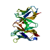 6dsiS S: Starting model for refinement C: citing same article ( |
|---|---|
| Similar structure data |
-Links
 PDBj
PDBj

- Assembly
| Deposited unit | 
| ||||||||||||||||||
|---|---|---|---|---|---|---|---|---|---|---|---|---|---|---|---|---|---|---|---|
| 1 | 
| ||||||||||||||||||
| 2 | 
| ||||||||||||||||||
| Unit cell |
| ||||||||||||||||||
| Noncrystallographic symmetry (NCS) | NCS domain:
NCS domain segments: Component-ID: 1 / Ens-ID: ens_1 / Beg auth comp-ID: GLU / Beg label comp-ID: GLU / End auth comp-ID: LYS / End label comp-ID: LYS / Auth seq-ID: 3 - 241 / Label seq-ID: 3 - 241
NCS oper: (Code: givenMatrix: (-0.999509173016, 0.0237495284529, 0.0204297076704), (-0.00856401377243, -0.834434178636, 0.551041068517), (0.0301342118752, 0.550595642394, 0.834228007113)Vector: 44. ...NCS oper: (Code: given Matrix: (-0.999509173016, 0.0237495284529, 0.0204297076704), Vector: |
-Components
| #1: Antibody | Mass: 27129.166 Da / Num. of mol.: 2 Source method: isolated from a genetically manipulated source Source: (gene. exp.) Homo sapiens (human) / Production host:  #2: Chemical | ChemComp-GOL / |   Mass: 92.094 Da / Num. of mol.: 1 / Source method: obtained synthetically / Formula: C3H8O3 Mass: 92.094 Da / Num. of mol.: 1 / Source method: obtained synthetically / Formula: C3H8O3#3: Water | ChemComp-HOH / |  Mass: 18.015 Da / Num. of mol.: 72 / Source method: isolated from a natural source / Formula: H2O Mass: 18.015 Da / Num. of mol.: 72 / Source method: isolated from a natural source / Formula: H2OHas ligand of interest | N | Has protein modification | Y | |
|---|
-Experimental details
-Experiment
| Experiment | Method: X-RAY DIFFRACTION / Number of used crystals: 1 |
|---|
- Sample preparation
| Crystal | Density Matthews: 3.93 Å3/Da / Density % sol: 68.73 % |
|---|---|
| Crystal grow | Temperature: 294 K / Method: vapor diffusion, hanging drop / Details: 4M Formate |
-Data collection
| Diffraction | Mean temperature: 100 K / Serial crystal experiment: N |
|---|---|
| Diffraction source | Source: SYNCHROTRON / Site: ESRF  / Beamline: ID30B / Wavelength: 0.97625 Å / Beamline: ID30B / Wavelength: 0.97625 Å |
| Detector | Type: DECTRIS PILATUS 6M / Detector: PIXEL / Date: Sep 28, 2021 |
| Radiation | Protocol: SINGLE WAVELENGTH / Monochromatic (M) / Laue (L): M / Scattering type: x-ray |
| Radiation wavelength | Wavelength: 0.97625 Å / Relative weight: 1 |
| Reflection | Resolution: 2.75→75.64 Å / Num. obs: 23027 / % possible obs: 99.6 % / Redundancy: 10 % / Biso Wilson estimate: 52.72 Å2 / CC1/2: 0.995 / Net I/σ(I): 12.9 |
| Reflection shell | Resolution: 2.75→2.8 Å / Mean I/σ(I) obs: 3.4 / Num. unique obs: 1110 / CC1/2: 0.84 |
- Processing
| Software |
| |||||||||||||||||||||||||||||||||||||||||||||||||||||||||||||||
|---|---|---|---|---|---|---|---|---|---|---|---|---|---|---|---|---|---|---|---|---|---|---|---|---|---|---|---|---|---|---|---|---|---|---|---|---|---|---|---|---|---|---|---|---|---|---|---|---|---|---|---|---|---|---|---|---|---|---|---|---|---|---|---|---|
| Refinement | Method to determine structure: MOLECULAR REPLACEMENT Starting model: 6DSI Resolution: 2.75→75.64 Å / SU ML: 0.2696 / Cross valid method: FREE R-VALUE / σ(F): 1.35 / Phase error: 20.2265 Stereochemistry target values: GeoStd + Monomer Library + CDL v1.2
| |||||||||||||||||||||||||||||||||||||||||||||||||||||||||||||||
| Solvent computation | Shrinkage radii: 0.9 Å / VDW probe radii: 1.1 Å / Solvent model: FLAT BULK SOLVENT MODEL | |||||||||||||||||||||||||||||||||||||||||||||||||||||||||||||||
| Displacement parameters | Biso mean: 47.46 Å2 | |||||||||||||||||||||||||||||||||||||||||||||||||||||||||||||||
| Refinement step | Cycle: LAST / Resolution: 2.75→75.64 Å
| |||||||||||||||||||||||||||||||||||||||||||||||||||||||||||||||
| Refine LS restraints |
| |||||||||||||||||||||||||||||||||||||||||||||||||||||||||||||||
| Refine LS restraints NCS | Type: Torsion NCS / Rms dev position: 0.558538674498 Å | |||||||||||||||||||||||||||||||||||||||||||||||||||||||||||||||
| LS refinement shell |
|
