Movie
Movie Controller

[English] 日本語
 Yorodumi
- PDB-8h10: Structure of SARS-CoV-1 Spike Protein with Engineered x1 Disulfid... -
+ Open data

- Basic information
| Entry | Database: PDB / ID: 8h10 | ||||||
|---|---|---|---|---|---|---|---|
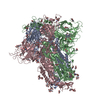
| Title | Structure of SARS-CoV-1 Spike Protein with Engineered x1 Disulfide (S370C and D967C), Locked-2 Conformation | ||||||
 Components Components | Spike glycoprotein | ||||||
 Keywords Keywords | VIRAL PROTEIN / PROTEIN ENGINEERING / SPIKE PROTEIN / SARS-COV-1 / SARS-COV | ||||||
| Function / homology |  Function and homology information Function and homology informationMaturation of spike protein / Translation of Structural Proteins / Virion Assembly and Release / Attachment and Entry / SARS-CoV-1 activates/modulates innate immune responses / symbiont-mediated-mediated suppression of host tetherin activity / positive regulation of viral entry into host cell / membrane fusion / host cell endoplasmic reticulum-Golgi intermediate compartment membrane / receptor-mediated virion attachment to host cell ...Maturation of spike protein / Translation of Structural Proteins / Virion Assembly and Release / Attachment and Entry / SARS-CoV-1 activates/modulates innate immune responses / symbiont-mediated-mediated suppression of host tetherin activity / positive regulation of viral entry into host cell / membrane fusion / host cell endoplasmic reticulum-Golgi intermediate compartment membrane / receptor-mediated virion attachment to host cell / host cell surface receptor binding / symbiont-mediated suppression of host innate immune response / endocytosis involved in viral entry into host cell / fusion of virus membrane with host plasma membrane / fusion of virus membrane with host endosome membrane / viral envelope / host cell plasma membrane / virion membrane / membrane / identical protein binding Similarity search - Function | ||||||
| Biological species |  Severe acute respiratory syndrome coronavirus Severe acute respiratory syndrome coronavirus | ||||||
| Method | ELECTRON MICROSCOPY / single particle reconstruction / cryo EM / Resolution: 2.99 Å | ||||||
 Authors Authors | Zhang, X. / Li, Z. / Liu, Y. / Wang, J. / Fu, L. / Wang, P. / He, J. / Xiong, X. | ||||||
| Funding support |  China, 1items China, 1items
| ||||||
 Citation Citation | Journal: Life Sci Alliance / Year: 2023 Title: Disulfide stabilization reveals conserved dynamic features between SARS-CoV-1 and SARS-CoV-2 spikes. Authors: Xixi Zhang / Zimu Li / Yanjun Zhang / Yutong Liu / Jingjing Wang / Banghui Liu / Qiuluan Chen / Qian Wang / Lutang Fu / Peiyi Wang / Xiaolin Zhong / Liang Jin / Qihong Yan / Ling Chen / Jun ...Authors: Xixi Zhang / Zimu Li / Yanjun Zhang / Yutong Liu / Jingjing Wang / Banghui Liu / Qiuluan Chen / Qian Wang / Lutang Fu / Peiyi Wang / Xiaolin Zhong / Liang Jin / Qihong Yan / Ling Chen / Jun He / Jincun Zhao / Xiaoli Xiong / Abstract: SARS-CoV-2 spike protein (S) is structurally dynamic and has been observed by cryo-EM to adopt a variety of prefusion conformations that can be categorized as locked, closed, and open. S-trimers ...SARS-CoV-2 spike protein (S) is structurally dynamic and has been observed by cryo-EM to adopt a variety of prefusion conformations that can be categorized as locked, closed, and open. S-trimers adopting locked conformations are tightly packed featuring structural elements incompatible with RBD in the "up" position. For SARS-CoV-2 S, it has been shown that the locked conformations are transient under neutral pH. Probably because of their transience, locked conformations remain largely uncharacterized for SARS-CoV-1 S. In this study, we introduced x1, x2, and x3 disulfides into SARS-CoV-1 S. Some of these disulfides have been shown to preserve rare locked conformations when introduced to SARS-CoV-2 S. Introduction of these disulfides allowed us to image a variety of locked and other rare conformations for SARS-CoV-1 S by cryo-EM. We identified bound cofactors and structural features that are associated with SARS-CoV-1 S locked conformations. We compare newly determined structures with other available spike structures of SARS-related CoVs to identify conserved features and discuss their possible functions. | ||||||
| History |
|
- Structure visualization
| Structure viewer | Molecule: MolmilJmol/JSmol |
|---|
- Downloads & links
-Download
| PDBx/mmCIF format | 8h10.cif.gz | 566.7 KB | Display | PDBx/mmCIF format |
|---|---|---|---|---|
| PDB format | pdb8h10.ent.gz | 456.6 KB | Display | PDB format |
| PDBx/mmJSON format | 8h10.json.gz | Tree view | PDBx/mmJSON format | |
| Others |  Other downloads Other downloads |
-Validation report
| Arichive directory | https://data.pdbj.org/pub/pdb/validation_reports/h1/8h10ftp://data.pdbj.org/pub/pdb/validation_reports/h1/8h10 | HTTPS FTP |
|---|
-Related structure data
| Related structure data |  34420MC  8h0xC  8h0yC  8h0zC 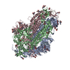 8h11C  8h12C 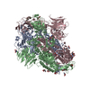 8h13C  8h14C 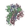 8h15C 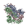 8h16C M: map data used to model this data C: citing same article ( |
|---|---|
| Similar structure data |
-Links
 PDBj
PDBj

- Assembly
| Deposited unit | 
|
|---|---|
| 1 |
|
-Components
| #1: Protein | Mass: 136679.359 Da / Num. of mol.: 3 / Mutation: S370C, D967C Source method: isolated from a genetically manipulated source Source: (gene. exp.) Severe acute respiratory syndrome coronavirusGene: S, 2 / Plasmid: pCDNA3.1 / Cell line (production host): Expi293 / Organ (production host): Kidney / Production host:  Homo sapiens (human) / References: UniProt: P59594 Homo sapiens (human) / References: UniProt: P59594#2: Polysaccharide | Source method: isolated from a genetically manipulated source #3: Sugar | ChemComp-NAG /   Type: D-saccharide, beta linking / Mass: 221.208 Da / Num. of mol.: 42 / Source method: obtained synthetically / Formula: C8H15NO6 Type: D-saccharide, beta linking / Mass: 221.208 Da / Num. of mol.: 42 / Source method: obtained synthetically / Formula: C8H15NO6#4: Chemical |   Mass: 582.646 Da / Num. of mol.: 3 / Source method: obtained synthetically / Formula: C33H34N4O6 Mass: 582.646 Da / Num. of mol.: 3 / Source method: obtained synthetically / Formula: C33H34N4O6#5: Chemical |   Mass: 280.445 Da / Num. of mol.: 3 / Source method: obtained synthetically / Formula: C18H32O2 Mass: 280.445 Da / Num. of mol.: 3 / Source method: obtained synthetically / Formula: C18H32O2Has ligand of interest | N | Has protein modification | Y | |
|---|
-Experimental details
-Experiment
| Experiment | Method: ELECTRON MICROSCOPY |
|---|---|
| EM experiment | Aggregation state: PARTICLE / 3D reconstruction method: single particle reconstruction |
- Sample preparation
| Component | Name: SARS-CoV-1 spike protein / Type: COMPLEX / Entity ID: #1 / Source: RECOMBINANT | ||||||||||||||||||||
|---|---|---|---|---|---|---|---|---|---|---|---|---|---|---|---|---|---|---|---|---|---|
| Molecular weight | Value: 0.41 MDa / Experimental value: NO | ||||||||||||||||||||
| Source (natural) | Organism: Severe acute respiratory syndrome coronavirus / Strain: SARS coronavirus Tor2 | ||||||||||||||||||||
| Source (recombinant) | Organism: Homo sapiens (human) / Cell: Expi293 / Plasmid: pCDNA3.1 | ||||||||||||||||||||
| Buffer solution | pH: 7.2 / Details: Gibco PBS C10010 | ||||||||||||||||||||
| Buffer component |
| ||||||||||||||||||||
| Specimen | Conc.: 4.7 mg/ml / Embedding applied: NO / Shadowing applied: NO / Staining applied: NO / Vitrification applied: YES | ||||||||||||||||||||
| Vitrification | Instrument: FEI VITROBOT MARK IV / Cryogen name: ETHANE / Humidity: 100 % / Chamber temperature: 295 K |
- Electron microscopy imaging
| Experimental equipment |  Model: Titan Krios / Image courtesy: FEI Company |
|---|---|
| Microscopy | Model: FEI TITAN KRIOS |
| Electron gun | Electron source:  FIELD EMISSION GUN / Accelerating voltage: 300 kV / Illumination mode: FLOOD BEAM FIELD EMISSION GUN / Accelerating voltage: 300 kV / Illumination mode: FLOOD BEAM |
| Electron lens | Mode: BRIGHT FIELD / Nominal magnification: 81000 X / Nominal defocus max: 2000 nm / Nominal defocus min: 800 nm / Cs: 2.7 mm / Alignment procedure: COMA FREE |
| Specimen holder | Cryogen: NITROGEN / Specimen holder model: FEI TITAN KRIOS AUTOGRID HOLDER |
| Image recording | Average exposure time: 2.37 sec. / Electron dose: 60 e/Å2 / Film or detector model: GATAN K3 BIOQUANTUM (6k x 4k) / Num. of grids imaged: 1 |
| EM imaging optics | Energyfilter name: GIF Bioquantum / Energyfilter slit width: 20 eV |
- Processing
| Software | Name: PHENIX / Version: 1.18.2_3874: / Classification: refinement | ||||||||||||||||||||||||||||||||||||||||||||||||||
|---|---|---|---|---|---|---|---|---|---|---|---|---|---|---|---|---|---|---|---|---|---|---|---|---|---|---|---|---|---|---|---|---|---|---|---|---|---|---|---|---|---|---|---|---|---|---|---|---|---|---|---|
| EM software |
| ||||||||||||||||||||||||||||||||||||||||||||||||||
| CTF correction | Type: PHASE FLIPPING AND AMPLITUDE CORRECTION | ||||||||||||||||||||||||||||||||||||||||||||||||||
| Particle selection | Num. of particles selected: 4604239 / Details: Particles were picked by Warp v1.09 | ||||||||||||||||||||||||||||||||||||||||||||||||||
| Symmetry | Point symmetry: C3 (3 fold cyclic) | ||||||||||||||||||||||||||||||||||||||||||||||||||
| 3D reconstruction | Resolution: 2.99 Å / Resolution method: FSC 0.143 CUT-OFF / Num. of particles: 22532 / Algorithm: FOURIER SPACE / Symmetry type: POINT | ||||||||||||||||||||||||||||||||||||||||||||||||||
| Atomic model building | B value: 55 / Protocol: FLEXIBLE FIT / Space: REAL | ||||||||||||||||||||||||||||||||||||||||||||||||||
| Atomic model building | PDB-ID: 7XU2 Accession code: 7XU2 / Source name: PDB / Type: experimental model | ||||||||||||||||||||||||||||||||||||||||||||||||||
| Refine LS restraints |
|