 Movie
Movie Controller

[English] 日本語
 Yorodumi
- PDB-8h0b: Structure of the thermolabile hemolysin from Vibrio alginolyticus... -
+ Open data

- Basic information
| Entry | Database: PDB / ID: 8h0b | ||||||
|---|---|---|---|---|---|---|---|
| Title | Structure of the thermolabile hemolysin from Vibrio alginolyticus (in complex with oleic acid) | ||||||
 Components Components | SGNH/GDSL hydrolase family protein | ||||||
 Keywords Keywords | HYDROLASE / Vibrio / phospholipase / SGNH hydrolase / GDSL lipase / transferase / thermolabile hemolysin | ||||||
| Function / homology |  Function and homology information Function and homology information | ||||||
| Biological species |  Vibrio alginolyticus (bacteria) Vibrio alginolyticus (bacteria) | ||||||
| Method |  X-RAY DIFFRACTION / SYNCHROTRON / MOLECULAR REPLACEMENT / Resolution: 1.931 Å X-RAY DIFFRACTION / SYNCHROTRON / MOLECULAR REPLACEMENT / Resolution: 1.931 Å | ||||||
 Authors Authors | Ma, Q. / Wang, C. | ||||||
| Funding support |  China, 1items China, 1items
| ||||||
 Citation Citation | Journal: Nat Commun / Year: 2023 Title: Catalytic site flexibility facilitates the substrate and catalytic promiscuity of Vibrio dual lipase/transferase. Authors: Wang, C. / Liu, C. / Zhu, X. / Peng, Q. / Ma, Q. | ||||||
| History |
|
- Structure visualization
| Structure viewer | Molecule: MolmilJmol/JSmol |
|---|
- Downloads & links
-Download
| PDBx/mmCIF format | 8h0b.cif.gz | 323.3 KB | Display | PDBx/mmCIF format |
|---|---|---|---|---|
| PDB format | pdb8h0b.ent.gz | 261.8 KB | Display | PDB format |
| PDBx/mmJSON format | 8h0b.json.gz | Tree view | PDBx/mmJSON format | |
| Others |  Other downloads Other downloads |
-Validation report
| Arichive directory | https://data.pdbj.org/pub/pdb/validation_reports/h0/8h0bftp://data.pdbj.org/pub/pdb/validation_reports/h0/8h0b | HTTPS FTP |
|---|
-Related structure data
| Related structure data |  8h09C 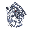 8h0aC  8h0cC 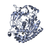 8h0dC  6jkzS S: Starting model for refinement C: citing same article ( |
|---|---|
| Similar structure data |
-Links
 PDBj
PDBj


- Assembly
| Deposited unit | 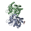
| ||||||||
|---|---|---|---|---|---|---|---|---|---|
| 1 | 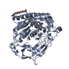
| ||||||||
| 2 | 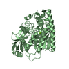
| ||||||||
| Unit cell |
|
-Components
| #1: Protein | Mass: 48378.746 Da / Num. of mol.: 2 Source method: isolated from a genetically manipulated source Source: (gene. exp.) Vibrio alginolyticus (bacteria) / Gene: F0254_14065 / Plasmid: pETM13 / Production host: #2: Chemical | ChemComp-OLA / |   Mass: 282.461 Da / Num. of mol.: 1 / Source method: obtained synthetically / Formula: C18H34O2 / Feature type: SUBJECT OF INVESTIGATION Mass: 282.461 Da / Num. of mol.: 1 / Source method: obtained synthetically / Formula: C18H34O2 / Feature type: SUBJECT OF INVESTIGATION#3: Chemical | ChemComp-1PE / |   Mass: 238.278 Da / Num. of mol.: 1 / Source method: obtained synthetically / Formula: C10H22O6 / Comment: precipitant*YM Mass: 238.278 Da / Num. of mol.: 1 / Source method: obtained synthetically / Formula: C10H22O6 / Comment: precipitant*YM#4: Chemical | ChemComp-MG / |   Mass: 24.305 Da / Num. of mol.: 1 / Source method: obtained synthetically / Formula: Mg Mass: 24.305 Da / Num. of mol.: 1 / Source method: obtained synthetically / Formula: Mg#5: Water | ChemComp-HOH / |  Mass: 18.015 Da / Num. of mol.: 201 / Source method: isolated from a natural source / Formula: H2O Mass: 18.015 Da / Num. of mol.: 201 / Source method: isolated from a natural source / Formula: H2OHas ligand of interest | Y | Has protein modification | Y | |
|---|
-Experimental details
-Experiment
| Experiment | Method: X-RAY DIFFRACTION / Number of used crystals: 1 |
|---|
- Sample preparation
| Crystal | Density Matthews: 1.99 Å3/Da / Density % sol: 38.21 % |
|---|---|
| Crystal grow | Temperature: 293 K / Method: vapor diffusion, sitting drop Details: The crystal of ValTLH in complex with oleic acid was grown in a drop containing 1.0 ul of protein solution (5 mg/ml in the buffer 10 mM HEPES pH 7.5, 150 mM NaCl, 1 mM DTT, 300 mM NDSB201 ...Details: The crystal of ValTLH in complex with oleic acid was grown in a drop containing 1.0 ul of protein solution (5 mg/ml in the buffer 10 mM HEPES pH 7.5, 150 mM NaCl, 1 mM DTT, 300 mM NDSB201 with 1 mM oleoyl coenzyme A lithium salt) and 1.0 ul of reservoir solution (22% polyethylene glycol 3350, 0.15 M magnesium formate). |
-Data collection
| Diffraction | Mean temperature: 100 K / Serial crystal experiment: N |
|---|---|
| Diffraction source | Source: SYNCHROTRON / Site: SSRF / Beamline: BL19U1 / Wavelength: 0.97852 Å |
| Detector | Type: DECTRIS PILATUS3 6M / Detector: PIXEL / Date: Oct 6, 2020 |
| Radiation | Protocol: SINGLE WAVELENGTH / Monochromatic (M) / Laue (L): M / Scattering type: x-ray |
| Radiation wavelength | Wavelength: 0.97852 Å / Relative weight: 1 |
| Reflection | Resolution: 1.931→56.301 Å / Num. obs: 57066 / % possible obs: 99.9 % / Redundancy: 6.7 % / CC1/2: 0.999 / Rpim(I) all: 0.02 / Rrim(I) all: 0.051 / Rsym value: 0.047 / Net I/σ(I): 20.2 |
| Reflection shell | Resolution: 1.931→1.964 Å / Redundancy: 6.8 % / Mean I/σ(I) obs: 2.5 / Num. unique obs: 2812 / CC1/2: 0.915 / Rpim(I) all: 0.244 / Rrim(I) all: 0.645 / Rsym value: 0.596 / % possible all: 100 |
- Processing
| Software |
| ||||||||||||||||||||||||||||||||||||||||||||||||||||||||||||||||||||||||||||||||||||||||||||||||||||||||||||
|---|---|---|---|---|---|---|---|---|---|---|---|---|---|---|---|---|---|---|---|---|---|---|---|---|---|---|---|---|---|---|---|---|---|---|---|---|---|---|---|---|---|---|---|---|---|---|---|---|---|---|---|---|---|---|---|---|---|---|---|---|---|---|---|---|---|---|---|---|---|---|---|---|---|---|---|---|---|---|---|---|---|---|---|---|---|---|---|---|---|---|---|---|---|---|---|---|---|---|---|---|---|---|---|---|---|---|---|---|---|
| Refinement | Method to determine structure: MOLECULAR REPLACEMENT Starting model: 6JKZ Resolution: 1.931→56.3 Å / Cor.coef. Fo:Fc: 0.947 / Cor.coef. Fo:Fc free: 0.937 / SU R Cruickshank DPI: 0.17 / Cross valid method: THROUGHOUT / σ(F): 0 / SU R Blow DPI: 0.165 / SU Rfree Blow DPI: 0.14 / SU Rfree Cruickshank DPI: 0.143
| ||||||||||||||||||||||||||||||||||||||||||||||||||||||||||||||||||||||||||||||||||||||||||||||||||||||||||||
| Displacement parameters | Biso max: 122.69 Å2 / Biso mean: 44.87 Å2 / Biso min: 23.45 Å2
| ||||||||||||||||||||||||||||||||||||||||||||||||||||||||||||||||||||||||||||||||||||||||||||||||||||||||||||
| Refine analyze | Luzzati coordinate error obs: 0.25 Å | ||||||||||||||||||||||||||||||||||||||||||||||||||||||||||||||||||||||||||||||||||||||||||||||||||||||||||||
| Refinement step | Cycle: final / Resolution: 1.931→56.3 Å
| ||||||||||||||||||||||||||||||||||||||||||||||||||||||||||||||||||||||||||||||||||||||||||||||||||||||||||||
| Refine LS restraints |
| ||||||||||||||||||||||||||||||||||||||||||||||||||||||||||||||||||||||||||||||||||||||||||||||||||||||||||||
| LS refinement shell | Resolution: 1.931→1.98 Å / Rfactor Rfree error: 0
| ||||||||||||||||||||||||||||||||||||||||||||||||||||||||||||||||||||||||||||||||||||||||||||||||||||||||||||
| Refinement TLS params. | Method: refined / Refine-ID: X-RAY DIFFRACTION
| ||||||||||||||||||||||||||||||||||||||||||||||||||||||||||||||||||||||||||||||||||||||||||||||||||||||||||||
| Refinement TLS group |
|
