 Movie
Movie Controller


+ Open data

- Basic information
| Entry | Database: PDB / ID: 8gqr | ||||||
|---|---|---|---|---|---|---|---|
| Title | Crystal structure of VioD with FAD | ||||||
 Components Components | VioD | ||||||
 Keywords Keywords | FLAVOPROTEIN / complex | ||||||
| Function / homology | : / FAD/NAD(P)-binding domain superfamily / oxidoreductase activity / nucleotide binding / FLAVIN-ADENINE DINUCLEOTIDE / VioD Function and homology information Function and homology information | ||||||
| Biological species |  Duganella sp. ZLP-XI (bacteria) Duganella sp. ZLP-XI (bacteria) | ||||||
| Method |  X-RAY DIFFRACTION / SYNCHROTRON / MOLECULAR REPLACEMENT / Resolution: 2.1 Å X-RAY DIFFRACTION / SYNCHROTRON / MOLECULAR REPLACEMENT / Resolution: 2.1 Å | ||||||
 Authors Authors | Ran, T. / Wang, W. / Xu, M. | ||||||
| Funding support |  China, 1items China, 1items
| ||||||
 Citation Citation | Journal: Proteins / Year: 2023 Title: Structural basis for substrate binding and catalytic mechanism of the key enzyme VioD in the violacein synthesis pathway. Authors: Xu, M. / Xu, D. / Gao, M. / Zhuang, X. / Wang, W. / Sun, B. / Ran, T. | ||||||
| History |
|
- Structure visualization
| Structure viewer | Molecule: MolmilJmol/JSmol |
|---|
- Downloads & links
-Download
| PDBx/mmCIF format | 8gqr.cif.gz | 174 KB | Display | PDBx/mmCIF format |
|---|---|---|---|---|
| PDB format | pdb8gqr.ent.gz | 132 KB | Display | PDB format |
| PDBx/mmJSON format | 8gqr.json.gz | Tree view | PDBx/mmJSON format | |
| Others |  Other downloads Other downloads |
-Validation report
| Arichive directory | https://data.pdbj.org/pub/pdb/validation_reports/gq/8gqrftp://data.pdbj.org/pub/pdb/validation_reports/gq/8gqr | HTTPS FTP |
|---|
-Related structure data
| Related structure data | 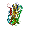 8h0mC 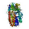 3c4aS S: Starting model for refinement C: citing same article ( |
|---|---|
| Similar structure data |
-Links
 PDBj
PDBj- Assembly
| Deposited unit | 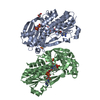
| ||||||||||||
|---|---|---|---|---|---|---|---|---|---|---|---|---|---|
| 1 | 
| ||||||||||||
| 2 | 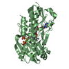
| ||||||||||||
| Unit cell |
|
-Components
| #1: Protein | Mass: 44543.488 Da / Num. of mol.: 2 Source method: isolated from a genetically manipulated source Source: (gene. exp.) Duganella sp. ZLP-XI (bacteria) / Production host: #2: Chemical | ChemComp-SO4 /   Mass: 96.063 Da / Num. of mol.: 6 / Source method: obtained synthetically / Formula: SO4 Mass: 96.063 Da / Num. of mol.: 6 / Source method: obtained synthetically / Formula: SO4#3: Chemical | ChemComp-GOL / |   Mass: 92.094 Da / Num. of mol.: 1 / Source method: obtained synthetically / Formula: C3H8O3 Mass: 92.094 Da / Num. of mol.: 1 / Source method: obtained synthetically / Formula: C3H8O3#4: Chemical |   Mass: 785.550 Da / Num. of mol.: 2 / Source method: obtained synthetically / Formula: C27H33N9O15P2 / Feature type: SUBJECT OF INVESTIGATION / Comment: FAD*YM Mass: 785.550 Da / Num. of mol.: 2 / Source method: obtained synthetically / Formula: C27H33N9O15P2 / Feature type: SUBJECT OF INVESTIGATION / Comment: FAD*YM#5: Water | ChemComp-HOH / |  Mass: 18.015 Da / Num. of mol.: 488 / Source method: isolated from a natural source / Formula: H2O Mass: 18.015 Da / Num. of mol.: 488 / Source method: isolated from a natural source / Formula: H2OHas ligand of interest | Y | |
|---|
-Experimental details
-Experiment
| Experiment | Method: X-RAY DIFFRACTION / Number of used crystals: 1 |
|---|
- Sample preparation
| Crystal | Density Matthews: 2.33 Å3/Da / Density % sol: 47.26 % |
|---|---|
| Crystal grow | Temperature: 295 K / Method: vapor diffusion, sitting drop / Details: PEG 3350 |
-Data collection
| Diffraction | Mean temperature: 100 K / Serial crystal experiment: N |
|---|---|
| Diffraction source | Source: SYNCHROTRON / Site: SSRF / Beamline: BL17U1 / Wavelength: 0.9791 Å |
| Detector | Type: ADSC QUANTUM 315r / Detector: CCD / Date: May 31, 2013 |
| Radiation | Protocol: SINGLE WAVELENGTH / Monochromatic (M) / Laue (L): M / Scattering type: x-ray |
| Radiation wavelength | Wavelength: 0.9791 Å / Relative weight: 1 |
| Reflection | Resolution: 2.1→19.77 Å / Num. obs: 49417 / % possible obs: 99.7 % / Redundancy: 7.3 % / CC1/2: 0.982 / Rpim(I) all: 0.13 / Net I/σ(I): 6.2 |
| Reflection shell | Resolution: 2.1→2.16 Å / Num. unique obs: 3951 / CC1/2: 0.499 / Rpim(I) all: 0.677 |
- Processing
| Software |
| ||||||||||||||||||||||||
|---|---|---|---|---|---|---|---|---|---|---|---|---|---|---|---|---|---|---|---|---|---|---|---|---|---|
| Refinement | Method to determine structure: MOLECULAR REPLACEMENT Starting model: 3C4A Resolution: 2.1→19.77 Å / Cross valid method: FREE R-VALUE Stereochemistry target values: GeoStd + Monomer Library + CDL v1.2
| ||||||||||||||||||||||||
| Displacement parameters | Biso mean: 26.75 Å2 | ||||||||||||||||||||||||
| Refinement step | Cycle: LAST / Resolution: 2.1→19.77 Å
| ||||||||||||||||||||||||
| Refine LS restraints |
|
