 Movie
Movie Controller


+ Open data

- Basic information
| Entry | Database: PDB / ID: 8gq4 | ||||||
|---|---|---|---|---|---|---|---|
| Title | Histone acetyltransferase Rtt109 mutant-N195A | ||||||
 Components Components | Histone acetyltransferase RTT109 | ||||||
 Keywords Keywords | TRANSFERASE / Histone acetyltransferase Rtt109 | ||||||
| Function / homology |  Function and homology information Function and homology informationregulation of phenotypic switching / negative regulation of filamentous growth of a population of unicellular organisms / filamentous growth of a population of unicellular organisms / histone H3K56 acetyltransferase activity / phenotypic switching / filamentous growth / histone H3 acetyltransferase activity / histone acetyltransferase / DNA damage response / regulation of DNA-templated transcription / nucleus Similarity search - Function | ||||||
| Biological species |  Candida albicans (yeast) Candida albicans (yeast) | ||||||
| Method |  X-RAY DIFFRACTION / SYNCHROTRON / MOLECULAR REPLACEMENT / Resolution: 1.77 Å X-RAY DIFFRACTION / SYNCHROTRON / MOLECULAR REPLACEMENT / Resolution: 1.77 Å | ||||||
 Authors Authors | Chen, Y.J. / Su, D. | ||||||
| Funding support |  China, 1items China, 1items
| ||||||
 Citation Citation | Journal: To Be Published Title: Structure of Histone acetyltransferase Rtt109 mutant-N195A Authors: Chen, Y.J. / Su, D. | ||||||
| History |
|
- Structure visualization
| Structure viewer | Molecule: MolmilJmol/JSmol |
|---|
- Downloads & links
-Download
| PDBx/mmCIF format | 8gq4.cif.gz | 102.3 KB | Display | PDBx/mmCIF format |
|---|---|---|---|---|
| PDB format | pdb8gq4.ent.gz | 61.5 KB | Display | PDB format |
| PDBx/mmJSON format | 8gq4.json.gz | Tree view | PDBx/mmJSON format | |
| Others |  Other downloads Other downloads |
-Validation report
| Summary document | 8gq4_validation.pdf.gz | 439.3 KB | Display | wwPDB validaton report |
|---|---|---|---|---|
| Full document | 8gq4_full_validation.pdf.gz | 444.8 KB | Display | |
| Data in XML | 8gq4_validation.xml.gz | 15.5 KB | Display | |
| Data in CIF | 8gq4_validation.cif.gz | 22 KB | Display | |
| Arichive directory | https://data.pdbj.org/pub/pdb/validation_reports/gq/8gq4ftp://data.pdbj.org/pub/pdb/validation_reports/gq/8gq4 | HTTPS FTP |
-Related structure data
| Related structure data | 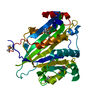 7bxwS S: Starting model for refinement |
|---|---|
| Similar structure data |
-Links
 PDBj
PDBj

- Assembly
| Deposited unit | 
| ||||||||||||
|---|---|---|---|---|---|---|---|---|---|---|---|---|---|
| 1 |
| ||||||||||||
| Unit cell |
|
-Components
| #1: Protein | Mass: 41866.422 Da / Num. of mol.: 1 / Mutation: N195A,S321L,S336L,S339L Source method: isolated from a genetically manipulated source Source: (gene. exp.) Candida albicans (yeast) / Strain: SC5314 / ATCC MYA-2876 / Gene: RTT109 / Production host:  |
|---|---|
| #2: Water | ChemComp-HOH /  Mass: 18.015 Da / Num. of mol.: 161 / Source method: isolated from a natural source / Formula: H2O Mass: 18.015 Da / Num. of mol.: 161 / Source method: isolated from a natural source / Formula: H2O |
| Has ligand of interest | Y |
| Has protein modification | Y |
-Experimental details
-Experiment
| Experiment | Method: X-RAY DIFFRACTION / Number of used crystals: 1 |
|---|
- Sample preparation
| Crystal | Density Matthews: 2.15 Å3/Da / Density % sol: 42.77 % |
|---|---|
| Crystal grow | Temperature: 291.15 K / Method: vapor diffusion, hanging drop / pH: 6 Details: 0.1 M Bis-Tris pH 6.0, 2% Tacsimate pH 6.0, 15~20% PEG 3350 |
-Data collection
| Diffraction | Mean temperature: 126.15 K / Serial crystal experiment: N |
|---|---|
| Diffraction source | Source: SYNCHROTRON / Site: SSRF / Beamline: BL17U1 / Wavelength: 0.9754 Å |
| Detector | Type: DECTRIS PILATUS3 6M / Detector: PIXEL / Date: Aug 3, 2017 |
| Radiation | Protocol: SINGLE WAVELENGTH / Monochromatic (M) / Laue (L): M / Scattering type: x-ray |
| Radiation wavelength | Wavelength: 0.9754 Å / Relative weight: 1 |
| Reflection | Resolution: 1.77→50 Å / Num. obs: 34445 / % possible obs: 100 % / Redundancy: 7.8 % / Biso Wilson estimate: 30.14 Å2 / Rmerge(I) obs: 0.185 / Net I/σ(I): 11.42 |
| Reflection shell | Resolution: 1.77→1.82 Å / Rmerge(I) obs: 0.335 / Num. unique obs: 2832 |
- Processing
| Software |
| |||||||||||||||||||||||||||||||||||||||||||||||||||||||||||||||||||||||||||||||||||||||||||
|---|---|---|---|---|---|---|---|---|---|---|---|---|---|---|---|---|---|---|---|---|---|---|---|---|---|---|---|---|---|---|---|---|---|---|---|---|---|---|---|---|---|---|---|---|---|---|---|---|---|---|---|---|---|---|---|---|---|---|---|---|---|---|---|---|---|---|---|---|---|---|---|---|---|---|---|---|---|---|---|---|---|---|---|---|---|---|---|---|---|---|---|---|
| Refinement | Method to determine structure: MOLECULAR REPLACEMENT Starting model: 7BXW Resolution: 1.77→49.78 Å / SU ML: 0.2031 / Cross valid method: FREE R-VALUE / σ(F): 1.39 / Phase error: 24.3364 Stereochemistry target values: GeoStd + Monomer Library + CDL v1.2
| |||||||||||||||||||||||||||||||||||||||||||||||||||||||||||||||||||||||||||||||||||||||||||
| Solvent computation | Shrinkage radii: 0.9 Å / VDW probe radii: 1.11 Å / Solvent model: FLAT BULK SOLVENT MODEL | |||||||||||||||||||||||||||||||||||||||||||||||||||||||||||||||||||||||||||||||||||||||||||
| Displacement parameters | Biso mean: 38.82 Å2 | |||||||||||||||||||||||||||||||||||||||||||||||||||||||||||||||||||||||||||||||||||||||||||
| Refinement step | Cycle: LAST / Resolution: 1.77→49.78 Å
| |||||||||||||||||||||||||||||||||||||||||||||||||||||||||||||||||||||||||||||||||||||||||||
| Refine LS restraints |
| |||||||||||||||||||||||||||||||||||||||||||||||||||||||||||||||||||||||||||||||||||||||||||
| LS refinement shell |
|
