 Movie
Movie Controller

[English] 日本語
 Yorodumi
- PDB-8gp3: Structure of beta-arrestin1 in complex with a phosphopeptide corr... -
+ Open data

- Basic information
| Entry | Database: PDB / ID: 8gp3 | ||||||
|---|---|---|---|---|---|---|---|
| Title | Structure of beta-arrestin1 in complex with a phosphopeptide corresponding to the human C-X-C chemokine receptor type 4, CXCR4 | ||||||
 Components Components |
| ||||||
 Keywords Keywords | SIGNALING PROTEIN / GPCR / Arrestin | ||||||
| Function / homology |  Function and homology information Function and homology informationV2 vasopressin receptor binding / alpha-1A adrenergic receptor binding / follicle-stimulating hormone receptor binding / TGFBR3 regulates TGF-beta signaling / G alpha (s) signalling events / sensory perception of touch / C-X-C motif chemokine 12 receptor activity / follicle-stimulating hormone signaling pathway / alpha-1B adrenergic receptor binding / protein phosphorylated amino acid binding ...V2 vasopressin receptor binding / alpha-1A adrenergic receptor binding / follicle-stimulating hormone receptor binding / TGFBR3 regulates TGF-beta signaling / G alpha (s) signalling events / sensory perception of touch / C-X-C motif chemokine 12 receptor activity / follicle-stimulating hormone signaling pathway / alpha-1B adrenergic receptor binding / protein phosphorylated amino acid binding / positive regulation of macrophage migration inhibitory factor signaling pathway / Lysosome Vesicle Biogenesis / myosin light chain binding / CXCL12-activated CXCR4 signaling pathway / Ub-specific processing proteases / Specification of primordial germ cells / angiotensin receptor binding / AP-2 adaptor complex binding / MAP2K and MAPK activation / Golgi Associated Vesicle Biogenesis / myelin maintenance / Developmental Lineage of Multipotent Pancreatic Progenitor Cells / C-X-C chemokine receptor activity / regulation of inositol trisphosphate biosynthetic process / positive regulation of vasculature development / negative regulation of GTPase activity / Cargo recognition for clathrin-mediated endocytosis / Clathrin-mediated endocytosis / Signaling by ROBO receptors / negative regulation of interleukin-8 production / clathrin-cargo adaptor activity / Formation of definitive endoderm / desensitization of G protein-coupled receptor signaling pathway / C-C chemokine binding / regulation of G protein-coupled receptor signaling pathway / C-C chemokine receptor activity / arrestin family protein binding / anchoring junction / G protein-coupled receptor internalization / mitogen-activated protein kinase kinase binding / Chemokine receptors bind chemokines / Thrombin signalling through proteinase activated receptors (PARs) / dendritic cell chemotaxis / clathrin binding / sensory perception / response to morphine / stress fiber assembly / negative regulation of interleukin-6 production / positive regulation of oligodendrocyte differentiation / cellular response to cytokine stimulus / positive regulation of Rho protein signal transduction / negative regulation of protein phosphorylation / cell leading edge / pseudopodium / negative regulation of Notch signaling pathway / phototransduction / positive regulation of receptor internalization / Binding and entry of HIV virion / regulation of cell adhesion / cysteine-type endopeptidase inhibitor activity / coreceptor activity / neurogenesis / insulin-like growth factor receptor binding / clathrin-coated pit / negative regulation of protein ubiquitination / intracellular glucose homeostasis / nuclear estrogen receptor binding / positive regulation of protein ubiquitination / positive regulation of insulin secretion involved in cellular response to glucose stimulus / GTPase activator activity / calcium-mediated signaling / cell chemotaxis / ubiquitin binding / brain development / phosphoprotein binding / negative regulation of ERK1 and ERK2 cascade / positive regulation of protein phosphorylation / response to virus / G protein-coupled receptor binding / G protein-coupled receptor activity / endocytosis / adenylate cyclase-modulating G protein-coupled receptor signaling pathway / adenylate cyclase-inhibiting G protein-coupled receptor signaling pathway / late endosome / positive regulation of cold-induced thermogenesis / protein transport / positive regulation of cytosolic calcium ion concentration / virus receptor activity / actin binding / cytoplasmic vesicle / regulation of apoptotic process / negative regulation of neuron apoptotic process / G alpha (i) signalling events / molecular adaptor activity / dendritic spine / basolateral plasma membrane / ubiquitin-dependent protein catabolic process / early endosome / proteasome-mediated ubiquitin-dependent protein catabolic process / response to hypoxia Similarity search - Function | ||||||
| Biological species |   Homo sapiens (human) Homo sapiens (human) | ||||||
| Method | ELECTRON MICROSCOPY / single particle reconstruction / cryo EM / Resolution: 4.8 Å | ||||||
 Authors Authors | Maharana, J. / Sarma, P. / Yadav, M.K. / Banerjee, R. / Shukla, A.K. | ||||||
| Funding support |  India, 1items India, 1items
| ||||||
 Citation Citation | Journal: Mol Cell / Year: 2023 Title: Structural snapshots uncover a key phosphorylation motif in GPCRs driving β-arrestin activation. Authors: Jagannath Maharana / Parishmita Sarma / Manish K Yadav / Sayantan Saha / Vinay Singh / Shirsha Saha / Mohamed Chami / Ramanuj Banerjee / Arun K Shukla /  Abstract: Agonist-induced GPCR phosphorylation is a key determinant for the binding and activation of β-arrestins (βarrs). However, it is not entirely clear how different GPCRs harboring divergent ...Agonist-induced GPCR phosphorylation is a key determinant for the binding and activation of β-arrestins (βarrs). However, it is not entirely clear how different GPCRs harboring divergent phosphorylation patterns impart converging active conformation on βarrs leading to broadly conserved functional responses such as desensitization, endocytosis, and signaling. Here, we present multiple cryo-EM structures of activated βarrs in complex with distinct phosphorylation patterns derived from the carboxyl terminus of different GPCRs. These structures help identify a P-X-P-P type phosphorylation motif in GPCRs that interacts with a spatially organized K-K-R-R-K-K sequence in the N-domain of βarrs. Sequence analysis of the human GPCRome reveals the presence of this phosphorylation pattern in a large number of receptors, and its contribution in βarr activation is demonstrated by targeted mutagenesis experiments combined with an intrabody-based conformational sensor. Taken together, our findings provide important structural insights into the ability of distinct GPCRs to activate βarrs through a significantly conserved mechanism. #1: Journal: Mol.Cell / Year: 2023Title: Structure of beta-arrestin in complex with a phosphopeptide Authors: Maharana, J. / Sarma, P. / Yadav, M.K. / Banerjee, R. / Shukla, A.K. | ||||||
| History |
|
- Structure visualization
| Structure viewer | Molecule: MolmilJmol/JSmol |
|---|
- Downloads & links
-Download
| PDBx/mmCIF format | 8gp3.cif.gz | 269.4 KB | Display | PDBx/mmCIF format |
|---|---|---|---|---|
| PDB format | pdb8gp3.ent.gz | 214.6 KB | Display | PDB format |
| PDBx/mmJSON format | 8gp3.json.gz | Tree view | PDBx/mmJSON format | |
| Others |  Other downloads Other downloads |
-Validation report
| Arichive directory | https://data.pdbj.org/pub/pdb/validation_reports/gp/8gp3ftp://data.pdbj.org/pub/pdb/validation_reports/gp/8gp3 | HTTPS FTP |
|---|
-Related structure data
| Related structure data | 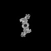 34188MC  8go8C  8gocC 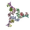 8gooC 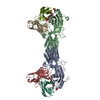 8i0nC 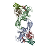 8i0qC 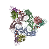 8i0zC 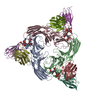 8i10C M: map data used to model this data C: citing same article ( |
|---|---|
| Similar structure data |


-Links
 PDBj
PDBj


- Assembly
| Deposited unit | 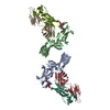
|
|---|---|
| 1 |
|
-Components
| #1: Protein | Mass: 47088.508 Da / Num. of mol.: 2 Source method: isolated from a genetically manipulated source Source: (gene. exp.)  #2: Protein/peptide | Mass: 2380.530 Da / Num. of mol.: 2 / Source method: obtained synthetically / Source: (synth.) Homo sapiens (human) / References: UniProt: P61073#3: Antibody | Mass: 25512.354 Da / Num. of mol.: 2 Source method: isolated from a genetically manipulated source Source: (gene. exp.) #4: Antibody | Mass: 23435.064 Da / Num. of mol.: 2 Source method: isolated from a genetically manipulated source Source: (gene. exp.) Has ligand of interest | Y | Has protein modification | Y | |
|---|
-Experimental details
-Experiment
| Experiment | Method: ELECTRON MICROSCOPY |
|---|---|
| EM experiment | Aggregation state: PARTICLE / 3D reconstruction method: single particle reconstruction |
- Sample preparation
| Component |
| ||||||||||||||||||||||||||||||
|---|---|---|---|---|---|---|---|---|---|---|---|---|---|---|---|---|---|---|---|---|---|---|---|---|---|---|---|---|---|---|---|
| Molecular weight | Value: 0.19 MDa / Experimental value: YES | ||||||||||||||||||||||||||||||
| Source (natural) |
| ||||||||||||||||||||||||||||||
| Source (recombinant) |
| ||||||||||||||||||||||||||||||
| Buffer solution | pH: 7.4 | ||||||||||||||||||||||||||||||
| Buffer component |
| ||||||||||||||||||||||||||||||
| Specimen | Embedding applied: NO / Shadowing applied: NO / Staining applied: NO / Vitrification applied: YES | ||||||||||||||||||||||||||||||
| Specimen support | Grid material: COPPER / Grid mesh size: 300 divisions/in. / Grid type: Quantifoil R2/2 | ||||||||||||||||||||||||||||||
| Vitrification | Instrument: LEICA EM GP / Cryogen name: ETHANE / Humidity: 90 % / Chamber temperature: 283.15 K / Details: Blotted for 3 seconds before plunging. |
- Electron microscopy imaging
| Microscopy | Model: TFS GLACIOS |
|---|---|
| Electron gun | Electron source:  FIELD EMISSION GUN / Accelerating voltage: 200 kV / Illumination mode: FLOOD BEAM FIELD EMISSION GUN / Accelerating voltage: 200 kV / Illumination mode: FLOOD BEAM |
| Electron lens | Mode: BRIGHT FIELD / Nominal magnification: 46000 X / Nominal defocus max: 2500 nm / Nominal defocus min: 500 nm / Cs: 2.7 mm / Alignment procedure: COMA FREE |
| Specimen holder | Cryogen: NITROGEN |
| Image recording | Electron dose: 49.3 e/Å2 / Detector mode: COUNTING / Film or detector model: GATAN K3 (6k x 4k) / Num. of real images: 5637 |
| Image scans | Movie frames/image: 40 |
- Processing
| EM software |
| ||||||||||||||||||||||||||||||||
|---|---|---|---|---|---|---|---|---|---|---|---|---|---|---|---|---|---|---|---|---|---|---|---|---|---|---|---|---|---|---|---|---|---|
| CTF correction | Type: NONE | ||||||||||||||||||||||||||||||||
| Particle selection | Num. of particles selected: 3236193 | ||||||||||||||||||||||||||||||||
| Symmetry | Point symmetry: C2 (2 fold cyclic) | ||||||||||||||||||||||||||||||||
| 3D reconstruction | Resolution: 4.8 Å / Resolution method: FSC 0.143 CUT-OFF / Num. of particles: 53387 / Symmetry type: POINT | ||||||||||||||||||||||||||||||||
| Atomic model building | Protocol: FLEXIBLE FIT / Space: REAL | ||||||||||||||||||||||||||||||||
| Atomic model building | PDB-ID: 8GO8 Accession code: 8GO8 / Source name: PDB / Type: experimental model |
