 Movie
Movie Controller


+ Open data

- Basic information
| Entry | Database: PDB / ID: 8gnk | |||||||||
|---|---|---|---|---|---|---|---|---|---|---|
| Title | CryoEM structure of cytosol-facing, substrate-bound ratGAT1 | |||||||||
 Components Components |
| |||||||||
 Keywords Keywords | MEMBRANE PROTEIN / Neurotransmitter sodium symporter / GABA transporter / solute carrier 6 / secondary active transport | |||||||||
| Function / homology |  Function and homology information Function and homology informationSLC-mediated transport of neurotransmitters / Reuptake of GABA / neurotransmitter reuptake / inorganic anion import across plasma membrane / sodium:chloride symporter activity / negative regulation of synaptic transmission, GABAergic / gamma-aminobutyric acid transmembrane transporter activity / gamma-aminobutyric acid import / gamma-aminobutyric acid:sodium:chloride symporter activity / response to methamphetamine hydrochloride ...SLC-mediated transport of neurotransmitters / Reuptake of GABA / neurotransmitter reuptake / inorganic anion import across plasma membrane / sodium:chloride symporter activity / negative regulation of synaptic transmission, GABAergic / gamma-aminobutyric acid transmembrane transporter activity / gamma-aminobutyric acid import / gamma-aminobutyric acid:sodium:chloride symporter activity / response to methamphetamine hydrochloride / amino acid:sodium symporter activity / response to sucrose / positive regulation of gamma-aminobutyric acid secretion / response to purine-containing compound / sodium ion import across plasma membrane / amino acid transport / associative learning / chloride transmembrane transport / learning / sodium ion transmembrane transport / response to cocaine / synapse organization / response to calcium ion / response to lead ion / GABA-ergic synapse / response to toxic substance / memory / response to estradiol / presynaptic membrane / postsynaptic membrane / response to xenobiotic stimulus / axon / neuronal cell body / cell surface / metal ion binding / identical protein binding / plasma membrane Similarity search - Function | |||||||||
| Biological species |  | |||||||||
| Method | ELECTRON MICROSCOPY / single particle reconstruction / cryo EM / Resolution: 3.1 Å | |||||||||
 Authors Authors | Nayak, S.R. / Joseph, D. / Penmatsa, A. | |||||||||
| Funding support |  India, 1items India, 1items
| |||||||||
 Citation Citation | Journal: Nat Struct Mol Biol / Year: 2023 Title: Cryo-EM structure of GABA transporter 1 reveals substrate recognition and transport mechanism. Authors: Smruti Ranjan Nayak / Deepthi Joseph / Georg Höfner / Archishman Dakua / Arunabh Athreya / Klaus T Wanner / Baruch I Kanner / Aravind Penmatsa /    Abstract: The inhibitory neurotransmitter γ-aminobutyric acid (GABA) is cleared from the synaptic cleft by the sodium- and chloride-coupled GABA transporter GAT1. Inhibition of GAT1 prolongs the GABAergic ...The inhibitory neurotransmitter γ-aminobutyric acid (GABA) is cleared from the synaptic cleft by the sodium- and chloride-coupled GABA transporter GAT1. Inhibition of GAT1 prolongs the GABAergic signaling at the synapse and is a strategy to treat certain forms of epilepsy. In this study, we present the cryo-electron microscopy structure of Rattus norvegicus GABA transporter 1 (rGAT1) at a resolution of 3.1 Å. The structure elucidation was facilitated by epitope transfer of a fragment-antigen binding (Fab) interaction site from the Drosophila dopamine transporter (dDAT) to rGAT1. The structure reveals rGAT1 in a cytosol-facing conformation, with a linear density in the primary binding site that accommodates a molecule of GABA, a displaced ion density proximal to Na site 1 and a bound chloride ion. A unique insertion in TM10 aids the formation of a compact, closed extracellular gate. Besides yielding mechanistic insights into ion and substrate recognition, our study will enable the rational design of specific antiepileptics. | |||||||||
| History |
|
- Structure visualization
| Structure viewer | Molecule: MolmilJmol/JSmol |
|---|
- Downloads & links
-Download
| PDBx/mmCIF format | 8gnk.cif.gz | 174.3 KB | Display | PDBx/mmCIF format |
|---|---|---|---|---|
| PDB format | pdb8gnk.ent.gz | 130.2 KB | Display | PDB format |
| PDBx/mmJSON format | 8gnk.json.gz | Tree view | PDBx/mmJSON format | |
| Others |  Other downloads Other downloads |
-Validation report
| Arichive directory | https://data.pdbj.org/pub/pdb/validation_reports/gn/8gnkftp://data.pdbj.org/pub/pdb/validation_reports/gn/8gnk | HTTPS FTP |
|---|
-Related structure data
| Related structure data |  34167MC M: map data used to model this data C: citing same article ( |
|---|---|
| Similar structure data |
-Links
 PDBj
PDBj


- Assembly
| Deposited unit | 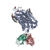
|
|---|---|
| 1 |
|
-Components
-Antibody , 2 types, 2 molecules LH
| #2: Antibody | Mass: 23306.586 Da / Num. of mol.: 1 Source method: isolated from a genetically manipulated source Source: (gene. exp.)   Spodoptera frugiperda (fall armyworm) Spodoptera frugiperda (fall armyworm) |
|---|---|
| #3: Antibody | Mass: 23619.430 Da / Num. of mol.: 1 Source method: isolated from a genetically manipulated source Source: (gene. exp.) Spodoptera frugiperda (fall armyworm) |
-Protein / Sugars , 2 types, 3 molecules A
| #1: Protein | Mass: 63200.676 Da / Num. of mol.: 1 Mutation: F312Y, Y481S, D482E, N483D, Q485R, E486D, V488I, S490F, R491P Source method: isolated from a genetically manipulated source Details: ratGAT1 construct / Source: (gene. exp.)  Homo sapiens (human) / References: UniProt: P23978 Homo sapiens (human) / References: UniProt: P23978 |
|---|---|
| #4: Sugar |  Type: D-saccharide, beta linking / Mass: 221.208 Da / Num. of mol.: 2 / Source method: obtained synthetically / Formula: C8H15NO6 Type: D-saccharide, beta linking / Mass: 221.208 Da / Num. of mol.: 2 / Source method: obtained synthetically / Formula: C8H15NO6 |
-Non-polymers , 6 types, 16 molecules 


| #5: Chemical | ChemComp-CL /  Mass: 35.453 Da / Num. of mol.: 1 / Source method: obtained synthetically / Formula: Cl Mass: 35.453 Da / Num. of mol.: 1 / Source method: obtained synthetically / Formula: Cl | ||||
|---|---|---|---|---|---|
| #6: Chemical | ChemComp-NA /  Mass: 22.990 Da / Num. of mol.: 1 / Source method: obtained synthetically / Formula: Na Mass: 22.990 Da / Num. of mol.: 1 / Source method: obtained synthetically / Formula: Na | ||||
| #7: Chemical | ChemComp-ABU /  Mass: 103.120 Da / Num. of mol.: 1 / Source method: obtained synthetically / Formula: C4H9NO2 / Feature type: SUBJECT OF INVESTIGATION / Comment: neurotransmitter, inhibitor*YM Mass: 103.120 Da / Num. of mol.: 1 / Source method: obtained synthetically / Formula: C4H9NO2 / Feature type: SUBJECT OF INVESTIGATION / Comment: neurotransmitter, inhibitor*YM | ||||
| #8: Chemical | ChemComp-CLR /  Mass: 386.654 Da / Num. of mol.: 6 / Source method: obtained synthetically / Formula: C27H46O Mass: 386.654 Da / Num. of mol.: 6 / Source method: obtained synthetically / Formula: C27H46O#9: Chemical | ChemComp-PTY / |  Mass: 734.039 Da / Num. of mol.: 1 / Source method: obtained synthetically / Formula: C40H80NO8P / Comment: phospholipid*YM Mass: 734.039 Da / Num. of mol.: 1 / Source method: obtained synthetically / Formula: C40H80NO8P / Comment: phospholipid*YM#10: Water | ChemComp-HOH / | Mass: 18.015 Da / Num. of mol.: 6 / Source method: isolated from a natural source / Formula: H2O |
-Details
| Has ligand of interest | Y |
|---|---|
| Has protein modification | Y |
-Experimental details
-Experiment
| Experiment | Method: ELECTRON MICROSCOPY |
|---|---|
| EM experiment | Aggregation state: PARTICLE / 3D reconstruction method: single particle reconstruction |
- Sample preparation
| Component |
| ||||||||||||||||||||||||||||||
|---|---|---|---|---|---|---|---|---|---|---|---|---|---|---|---|---|---|---|---|---|---|---|---|---|---|---|---|---|---|---|---|
| Molecular weight | Value: 111 kDa/nm / Experimental value: NO | ||||||||||||||||||||||||||||||
| Source (natural) |
| ||||||||||||||||||||||||||||||
| Source (recombinant) |
| ||||||||||||||||||||||||||||||
| Buffer solution | pH: 8 | ||||||||||||||||||||||||||||||
| Buffer component |
| ||||||||||||||||||||||||||||||
| Specimen | Conc.: 3.5 mg/ml / Embedding applied: NO / Shadowing applied: NO / Staining applied: NO / Vitrification applied: YES Details: The sample was a one to one complex of ratGAT1 with an antibody fragment and was homogenous. | ||||||||||||||||||||||||||||||
| Specimen support | Grid material: GOLD / Grid mesh size: 300 divisions/in. / Grid type: Quantifoil R0.6/1 | ||||||||||||||||||||||||||||||
| Vitrification | Cryogen name: ETHANE |
- Electron microscopy imaging
| Experimental equipment |  Model: Titan Krios / Image courtesy: FEI Company |
|---|---|
| Microscopy | Model: FEI TITAN KRIOS |
| Electron gun | Electron source:  FIELD EMISSION GUN / Accelerating voltage: 300 kV / Illumination mode: OTHER FIELD EMISSION GUN / Accelerating voltage: 300 kV / Illumination mode: OTHER |
| Electron lens | Mode: BRIGHT FIELD / Nominal magnification: 105000 X / Calibrated magnification: 105000 X / Nominal defocus max: 4000 nm / Nominal defocus min: 500 nm / Cs: 2.7 mm / C2 aperture diameter: 70 µm / Alignment procedure: BASIC |
| Specimen holder | Cryogen: NITROGEN / Specimen holder model: FEI TITAN KRIOS AUTOGRID HOLDER / Temperature (max): 100 K / Temperature (min): 100 K |
| Image recording | Average exposure time: 2.7 sec. / Electron dose: 50.09 e/Å2 / Film or detector model: GATAN K3 BIOQUANTUM (6k x 4k) / Num. of grids imaged: 1 / Num. of real images: 18152 Details: Images were collected in movie mode at 50 frames per image |
| EM imaging optics | Energyfilter name: GIF Bioquantum / Chromatic aberration corrector: No applicable / Energyfilter slit width: 20 eV / Phase plate: OTHER / Spherical aberration corrector: Not applicable |
- Processing
| Software | Name: PHENIX / Version: 1.20rc4_4425: / Classification: refinement | ||||||||||||||||||||||||||||||||||||||||
|---|---|---|---|---|---|---|---|---|---|---|---|---|---|---|---|---|---|---|---|---|---|---|---|---|---|---|---|---|---|---|---|---|---|---|---|---|---|---|---|---|---|
| EM software |
| ||||||||||||||||||||||||||||||||||||||||
| CTF correction | Type: PHASE FLIPPING AND AMPLITUDE CORRECTION | ||||||||||||||||||||||||||||||||||||||||
| Particle selection | Num. of particles selected: 5838634 | ||||||||||||||||||||||||||||||||||||||||
| Symmetry | Point symmetry: C1 (asymmetric) | ||||||||||||||||||||||||||||||||||||||||
| 3D reconstruction | Resolution: 3.1 Å / Resolution method: FSC 0.143 CUT-OFF / Num. of particles: 872611 / Num. of class averages: 3 / Symmetry type: POINT | ||||||||||||||||||||||||||||||||||||||||
| Atomic model building | B value: 165 / Protocol: OTHER / Space: REAL / Target criteria: Correlation coefficient | ||||||||||||||||||||||||||||||||||||||||
| Refine LS restraints |
|
