 Movie
Movie Controller


+ Open data

- Basic information
| Entry | Database: PDB / ID: 8gmu | ||||||
|---|---|---|---|---|---|---|---|
| Title | Structure of lambda repressor in complex with RecA filament | ||||||
 Components Components |
| ||||||
 Keywords Keywords | DNA BINDING PROTEIN/DNA / SOS response / RecA / lambda repressor / Filament / DNA repair / Helical reconstruction / DNA BINDING PROTEIN-DNA COMPLEX | ||||||
| Function / homology |  Function and homology information Function and homology informationmaintenance of viral latency / DNA polymerase V complex / latency-replication decision / positive regulation of viral transcription / homologous recombination / negative regulation of transcription by competitive promoter binding / SOS response / recombinational repair / ATP-dependent DNA damage sensor activity / response to ionizing radiation ...maintenance of viral latency / DNA polymerase V complex / latency-replication decision / positive regulation of viral transcription / homologous recombination / negative regulation of transcription by competitive promoter binding / SOS response / recombinational repair / ATP-dependent DNA damage sensor activity / response to ionizing radiation / ATP-dependent activity, acting on DNA / translesion synthesis / core promoter sequence-specific DNA binding / cell motility / single-stranded DNA binding / DNA recombination / DNA-binding transcription factor binding / damaged DNA binding / DNA damage response / ATP hydrolysis activity / ATP binding / identical protein binding / cytoplasm Similarity search - Function | ||||||
| Biological species |  Escherichia phage Lambda (virus) Escherichia phage Lambda (virus) | ||||||
| Method | ELECTRON MICROSCOPY / helical reconstruction / cryo EM / Resolution: 2.78 Å | ||||||
 Authors Authors | Gao, B. / Feng, Y. | ||||||
| Funding support |  China, 1items China, 1items
| ||||||
 Citation Citation | Journal: Proc Natl Acad Sci U S A / Year: 2023 Title: Structural basis for regulation of SOS response in bacteria. Authors: Bo Gao / Liang Liang / Lu Su / Aijia Wen / Chun Zhou / Yu Feng / Abstract: In response to DNA damage, bacterial RecA protein forms filaments with the assistance of DinI protein. The RecA filaments stimulate the autocleavage of LexA, the repressor of more than 50 SOS genes, ...In response to DNA damage, bacterial RecA protein forms filaments with the assistance of DinI protein. The RecA filaments stimulate the autocleavage of LexA, the repressor of more than 50 SOS genes, and activate the SOS response. During the late phase of SOS response, the RecA filaments stimulate the autocleavage of UmuD and λ repressor CI, leading to mutagenic repair and lytic cycle, respectively. Here, we determined the cryo-electron microscopy structures of RecA filaments in complex with DinI, LexA, UmuD, and λCI by helical reconstruction. The structures reveal that LexA and UmuD dimers bind in the filament groove and cleave in an intramolecular and an intermolecular manner, respectively, while λCI binds deeply in the filament groove as a monomer. Despite their distinct folds and oligomeric states, all RecA filament binders recognize the same conserved protein features in the filament groove. The SOS response in bacteria can lead to mutagenesis and antimicrobial resistance, and our study paves the way for rational drug design targeting the bacterial SOS response. | ||||||
| History |
|
- Structure visualization
| Structure viewer | Molecule: MolmilJmol/JSmol |
|---|
- Downloads & links
-Download
| PDBx/mmCIF format | 8gmu.cif.gz | 163.6 KB | Display | PDBx/mmCIF format |
|---|---|---|---|---|
| PDB format | pdb8gmu.ent.gz | 126.7 KB | Display | PDB format |
| PDBx/mmJSON format | 8gmu.json.gz | Tree view | PDBx/mmJSON format | |
| Others |  Other downloads Other downloads |
-Validation report
| Summary document | 8gmu_validation.pdf.gz | 1.3 MB | Display | wwPDB validaton report |
|---|---|---|---|---|
| Full document | 8gmu_full_validation.pdf.gz | 1.3 MB | Display | |
| Data in XML | 8gmu_validation.xml.gz | 32.4 KB | Display | |
| Data in CIF | 8gmu_validation.cif.gz | 46.6 KB | Display | |
| Arichive directory | https://data.pdbj.org/pub/pdb/validation_reports/gm/8gmuftp://data.pdbj.org/pub/pdb/validation_reports/gm/8gmu | HTTPS FTP |
-Related structure data
| Related structure data |  34154MC  7ywaC 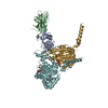 8gmsC 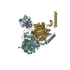 8gmtC M: map data used to model this data C: citing same article ( |
|---|---|
| Similar structure data |

-Links
 PDBj
PDBj


- Assembly
| Deposited unit | 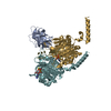
|
|---|---|
| 1 |
|
-Components
| #1: Protein | Mass: 14999.833 Da / Num. of mol.: 1 / Mutation: K192A Source method: isolated from a genetically manipulated source Source: (gene. exp.) Escherichia phage Lambda (virus) / Gene: cI, lambdap88 / Production host: | ||||||||
|---|---|---|---|---|---|---|---|---|---|
| #2: Protein | Mass: 38016.277 Da / Num. of mol.: 2 Source method: isolated from a genetically manipulated source Source: (gene. exp.) #3: DNA chain | | Mass: 1780.199 Da / Num. of mol.: 1 / Source method: obtained synthetically / Source: (synth.) #4: Chemical |   Mass: 24.305 Da / Num. of mol.: 2 / Source method: obtained synthetically / Formula: Mg Mass: 24.305 Da / Num. of mol.: 2 / Source method: obtained synthetically / Formula: Mg#5: Chemical |   Mass: 523.247 Da / Num. of mol.: 2 / Source method: obtained synthetically / Formula: C10H16N5O12P3S / Comment: ATP-gamma-S, energy-carrying molecule analogue*YM Mass: 523.247 Da / Num. of mol.: 2 / Source method: obtained synthetically / Formula: C10H16N5O12P3S / Comment: ATP-gamma-S, energy-carrying molecule analogue*YMHas ligand of interest | N | |
-Experimental details
-Experiment
| Experiment | Method: ELECTRON MICROSCOPY |
|---|---|
| EM experiment | Aggregation state: HELICAL ARRAY / 3D reconstruction method: helical reconstruction |
- Sample preparation
| Component | Name: RecA-CI complex / Type: COMPLEX / Entity ID: #1-#3 / Source: RECOMBINANT |
|---|---|
| Molecular weight | Experimental value: NO |
| Source (natural) | Organism: Escherichia phage Lambda (virus) |
| Source (recombinant) | Organism: |
| Buffer solution | pH: 7.5 |
| Specimen | Embedding applied: NO / Shadowing applied: NO / Staining applied: NO / Vitrification applied: YES |
| Vitrification | Cryogen name: ETHANE |
- Electron microscopy imaging
| Experimental equipment |  Model: Titan Krios / Image courtesy: FEI Company |
|---|---|
| Microscopy | Model: FEI TITAN KRIOS |
| Electron gun | Electron source:  FIELD EMISSION GUN / Accelerating voltage: 300 kV / Illumination mode: FLOOD BEAM FIELD EMISSION GUN / Accelerating voltage: 300 kV / Illumination mode: FLOOD BEAM |
| Electron lens | Mode: BRIGHT FIELD / Nominal defocus max: 1700 nm / Nominal defocus min: 800 nm |
| Image recording | Electron dose: 50 e/Å2 / Film or detector model: FEI FALCON IV (4k x 4k) |
- Processing
| CTF correction | Type: PHASE FLIPPING AND AMPLITUDE CORRECTION |
|---|---|
| Helical symmerty | Angular rotation/subunit: 118.1 ° / Axial rise/subunit: 31.5 Å / Axial symmetry: C1 |
| 3D reconstruction | Resolution: 2.78 Å / Resolution method: FSC 0.143 CUT-OFF / Num. of particles: 500031 / Symmetry type: HELICAL |
