[English] 日本語
 Yorodumi
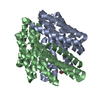
- PDB-8glt: Backbone model of de novo-designed chlorophyll-binding nanocage O32-15 -
+ Open data
- Basic information
| Entry | Database: PDB / ID: 8glt | |||||||||
|---|---|---|---|---|---|---|---|---|---|---|
| Title | Backbone model of de novo-designed chlorophyll-binding nanocage O32-15 | |||||||||
 Components Components |
| |||||||||
 Keywords Keywords | DE NOVO PROTEIN / nanocage / helical repeats / chlorophyll-binding / octahedral symmetry | |||||||||
| Biological species | synthetic construct (others) | |||||||||
| Method | ELECTRON MICROSCOPY / single particle reconstruction / cryo EM / Resolution: 6.5 Å | |||||||||
 Authors Authors | Redler, R.L. / Ennist, N.M. / Wang, S. / Baker, D. / Ekiert, D.C. / Bhabha, G. | |||||||||
| Funding support |  United States, 2items United States, 2items
| |||||||||
 Citation Citation |  Journal: Nat Chem Biol / Year: 2024 Journal: Nat Chem Biol / Year: 2024Title: De novo design of proteins housing excitonically coupled chlorophyll special pairs. Authors: Nathan M Ennist / Shunzhi Wang / Madison A Kennedy / Mariano Curti / George A Sutherland / Cvetelin Vasilev / Rachel L Redler / Valentin Maffeis / Saeed Shareef / Anthony V Sica / Ash Sueh ...Authors: Nathan M Ennist / Shunzhi Wang / Madison A Kennedy / Mariano Curti / George A Sutherland / Cvetelin Vasilev / Rachel L Redler / Valentin Maffeis / Saeed Shareef / Anthony V Sica / Ash Sueh Hua / Arundhati P Deshmukh / Adam P Moyer / Derrick R Hicks / Avi Z Swartz / Ralph A Cacho / Naia Novy / Asim K Bera / Alex Kang / Banumathi Sankaran / Matthew P Johnson / Amala Phadkule / Mike Reppert / Damian Ekiert / Gira Bhabha / Lance Stewart / Justin R Caram / Barry L Stoddard / Elisabet Romero / C Neil Hunter / David Baker /    Abstract: Natural photosystems couple light harvesting to charge separation using a 'special pair' of chlorophyll molecules that accepts excitation energy from the antenna and initiates an electron-transfer ...Natural photosystems couple light harvesting to charge separation using a 'special pair' of chlorophyll molecules that accepts excitation energy from the antenna and initiates an electron-transfer cascade. To investigate the photophysics of special pairs independently of the complexities of native photosynthetic proteins, and as a first step toward creating synthetic photosystems for new energy conversion technologies, we designed C-symmetric proteins that hold two chlorophyll molecules in closely juxtaposed arrangements. X-ray crystallography confirmed that one designed protein binds two chlorophylls in the same orientation as native special pairs, whereas a second designed protein positions them in a previously unseen geometry. Spectroscopy revealed that the chlorophylls are excitonically coupled, and fluorescence lifetime imaging demonstrated energy transfer. The cryo-electron microscopy structure of a designed 24-chlorophyll octahedral nanocage with a special pair on each edge closely matched the design model. The results suggest that the de novo design of artificial photosynthetic systems is within reach of current computational methods. | |||||||||
| History |
|
- Structure visualization
| Structure viewer | Molecule:  Molmil Molmil Jmol/JSmol Jmol/JSmol |
|---|
- Downloads & links
- Download
| PDBx/mmCIF format |  8glt.cif.gz 8glt.cif.gz | 1.9 MB | Display |  PDBx/mmCIF format PDBx/mmCIF format |
|---|---|---|---|---|
| PDB format |  pdb8glt.ent.gz pdb8glt.ent.gz | Display |  PDB format PDB format | |
| PDBx/mmJSON format |  8glt.json.gz 8glt.json.gz | Tree view |  PDBx/mmJSON format PDBx/mmJSON format | |
| Others |  Other downloads Other downloads |
-Validation report
| Summary document |  8glt_validation.pdf.gz 8glt_validation.pdf.gz | 1.6 MB | Display |  wwPDB validaton report wwPDB validaton report |
|---|---|---|---|---|
| Full document |  8glt_full_validation.pdf.gz 8glt_full_validation.pdf.gz | 1.7 MB | Display | |
| Data in XML |  8glt_validation.xml.gz 8glt_validation.xml.gz | 246.2 KB | Display | |
| Data in CIF |  8glt_validation.cif.gz 8glt_validation.cif.gz | 395.6 KB | Display | |
| Arichive directory |  https://data.pdbj.org/pub/pdb/validation_reports/gl/8glt https://data.pdbj.org/pub/pdb/validation_reports/gl/8glt ftp://data.pdbj.org/pub/pdb/validation_reports/gl/8glt ftp://data.pdbj.org/pub/pdb/validation_reports/gl/8glt | HTTPS FTP |
-Related structure data
| Related structure data |  40208MC  7unhC  7uniC  7unjC  8evmC M: map data used to model this data C: citing same article ( |
|---|
- Links
Links
- Assembly
| Deposited unit | 
|
|---|---|
| 1 |
|
- Components
| #1: Protein | Mass: 35889.559 Da / Num. of mol.: 24 Source method: isolated from a genetically manipulated source Source: (gene. exp.) synthetic construct (others) / Production host:  #2: Protein | Mass: 27799.957 Da / Num. of mol.: 24 Source method: isolated from a genetically manipulated source Source: (gene. exp.) synthetic construct (others) / Production host:  |
|---|
-Experimental details
-Experiment
| Experiment | Method: ELECTRON MICROSCOPY |
|---|---|
| EM experiment | Aggregation state: PARTICLE / 3D reconstruction method: single particle reconstruction |
- Sample preparation
| Component | Name: Chlorophyll-binding nanocage O32-15 loaded with ZnPPaM Type: COMPLEX / Entity ID: all / Source: RECOMBINANT |
|---|---|
| Molecular weight | Experimental value: NO |
| Source (natural) | Organism: synthetic construct (others) |
| Source (recombinant) | Organism:  |
| Buffer solution | pH: 8 |
| Specimen | Conc.: 0.7 mg/ml / Embedding applied: NO / Shadowing applied: NO / Staining applied: NO / Vitrification applied: YES |
| Specimen support | Grid material: COPPER / Grid mesh size: 300 divisions/in. / Grid type: Quantifoil R2/2 |
| Vitrification | Instrument: FEI VITROBOT MARK IV / Cryogen name: ETHANE / Humidity: 100 % / Chamber temperature: 295 K |
- Electron microscopy imaging
| Experimental equipment |  Model: Titan Krios / Image courtesy: FEI Company |
|---|---|
| Microscopy | Model: FEI TITAN KRIOS |
| Electron gun | Electron source:  FIELD EMISSION GUN / Accelerating voltage: 300 kV / Illumination mode: FLOOD BEAM FIELD EMISSION GUN / Accelerating voltage: 300 kV / Illumination mode: FLOOD BEAM |
| Electron lens | Mode: BRIGHT FIELD / Nominal magnification: 64000 X / Nominal defocus max: 2500 nm / Nominal defocus min: 300 nm |
| Specimen holder | Specimen holder model: FEI TITAN KRIOS AUTOGRID HOLDER |
| Image recording | Average exposure time: 2 sec. / Electron dose: 49.99 e/Å2 / Film or detector model: GATAN K3 BIOQUANTUM (6k x 4k) / Num. of grids imaged: 2 |
| EM imaging optics | Energyfilter slit width: 20 eV |
- Processing
| Software |
| ||||||||||||||||||||||||||||||||||||||||||||
|---|---|---|---|---|---|---|---|---|---|---|---|---|---|---|---|---|---|---|---|---|---|---|---|---|---|---|---|---|---|---|---|---|---|---|---|---|---|---|---|---|---|---|---|---|---|
| EM software |
| ||||||||||||||||||||||||||||||||||||||||||||
| CTF correction | Details: Patch CTF estimation / Type: PHASE FLIPPING AND AMPLITUDE CORRECTION | ||||||||||||||||||||||||||||||||||||||||||||
| Particle selection | Num. of particles selected: 359331 | ||||||||||||||||||||||||||||||||||||||||||||
| Symmetry | Point symmetry: O (octahedral) | ||||||||||||||||||||||||||||||||||||||||||||
| 3D reconstruction | Resolution: 6.5 Å / Resolution method: FSC 0.143 CUT-OFF / Num. of particles: 205014 / Symmetry type: POINT | ||||||||||||||||||||||||||||||||||||||||||||
| Atomic model building | Protocol: FLEXIBLE FIT / Space: REAL | ||||||||||||||||||||||||||||||||||||||||||||
| Refinement | Cross valid method: NONE Stereochemistry target values: GeoStd + Monomer Library + CDL v1.2 | ||||||||||||||||||||||||||||||||||||||||||||
| Displacement parameters | Biso mean: 0 Å2 | ||||||||||||||||||||||||||||||||||||||||||||
| Refine LS restraints |
|
 Movie
Movie Controller


 PDBj
PDBj
