[English] 日本語
Yorodumi
- PDB-7uni: De novo designed chlorophyll dimer protein with Zn pheophorbide a... -

+
Open data


ID or keywords:

Loading...

-
Basic information

Entry
Database: PDB / ID: 7uni
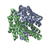
TitleDe novo designed chlorophyll dimer protein with Zn pheophorbide a methyl ester, SP2-ZnPPaM
ComponentsSP2-ZnPPaM designed chlorophyll dimer protein
KeywordsDE NOVO PROTEIN / design / homodimer / rosetta / symmetric / designed / circular tandem repeat protein / cTRP / chlorophyll / Zn pheophorbide a methyl ester / Zn
Function / homologyChem-OE9 / TRIETHYLENE GLYCOL / PHOSPHATE ION
Function and homology information
Biological speciessynthetic construct (others)
MethodX-RAY DIFFRACTION / SYNCHROTRON / MOLECULAR REPLACEMENT / Resolution: 2.5 Å
AuthorsKennedy, M.A. / Stoddard, B.L. / Ennist, N.M.
Funding support United States, 3items
OrganizationGrant numberCountry
National Science Foundation (NSF, United States)DGE-1762114 United States
Advanced Research Projects Agency-Energy (ARPA-E)DE-AR0001543
Audacious Project at the Institute for Protein Design68-2492: Audacious GC5: Next-Generation Nanoengineering
CitationJournal: Nat Chem Biol / Year: 2024
Title: De novo design of proteins housing excitonically coupled chlorophyll special pairs.
Authors: Nathan M Ennist / Shunzhi Wang / Madison A Kennedy / Mariano Curti / George A Sutherland / Cvetelin Vasilev / Rachel L Redler / Valentin Maffeis / Saeed Shareef / Anthony V Sica / Ash Sueh ...Authors: Nathan M Ennist / Shunzhi Wang / Madison A Kennedy / Mariano Curti / George A Sutherland / Cvetelin Vasilev / Rachel L Redler / Valentin Maffeis / Saeed Shareef / Anthony V Sica / Ash Sueh Hua / Arundhati P Deshmukh / Adam P Moyer / Derrick R Hicks / Avi Z Swartz / Ralph A Cacho / Nathan Novy / Asim K Bera / Alex Kang / Banumathi Sankaran / Matthew P Johnson / Amala Phadkule / Mike Reppert / Damian Ekiert / Gira Bhabha / Lance Stewart / Justin R Caram / Barry L Stoddard / Elisabet Romero / C Neil Hunter / David Baker /
Abstract: Natural photosystems couple light harvesting to charge separation using a 'special pair' of chlorophyll molecules that accepts excitation energy from the antenna and initiates an electron-transfer ...Natural photosystems couple light harvesting to charge separation using a 'special pair' of chlorophyll molecules that accepts excitation energy from the antenna and initiates an electron-transfer cascade. To investigate the photophysics of special pairs independently of the complexities of native photosynthetic proteins, and as a first step toward creating synthetic photosystems for new energy conversion technologies, we designed C-symmetric proteins that hold two chlorophyll molecules in closely juxtaposed arrangements. X-ray crystallography confirmed that one designed protein binds two chlorophylls in the same orientation as native special pairs, whereas a second designed protein positions them in a previously unseen geometry. Spectroscopy revealed that the chlorophylls are excitonically coupled, and fluorescence lifetime imaging demonstrated energy transfer. The cryo-electron microscopy structure of a designed 24-chlorophyll octahedral nanocage with a special pair on each edge closely matched the design model. The results suggest that the de novo design of artificial photosynthetic systems is within reach of current computational methods.
History
DepositionApr 11, 2022Deposition site: RCSB / Processing site: RCSB
Revision 1.0Apr 19, 2023Provider: repository / Type: Initial release
Revision 1.1Apr 3, 2024Group: Data collection / Refinement description
Category: chem_comp_atom / chem_comp_bond / pdbx_initial_refinement_model
Revision 1.2Jun 12, 2024Group: Database references / Category: citation / citation_author
Item: _citation.country / _citation.journal_abbrev ..._citation.country / _citation.journal_abbrev / _citation.journal_id_CSD / _citation.journal_id_ISSN / _citation.pdbx_database_id_DOI / _citation.pdbx_database_id_PubMed / _citation.title / _citation.year

-
Structure visualization

Structure viewerMolecule:
MolmilJmol/JSmol

Downloads & links

-
Assembly

Deposited unit
A: SP2-ZnPPaM designed chlorophyll dimer protein
C: SP2-ZnPPaM designed chlorophyll dimer protein
B: SP2-ZnPPaM designed chlorophyll dimer protein
D: SP2-ZnPPaM designed chlorophyll dimer protein
hetero molecules


Theoretical massNumber of molelcules
Total (without water)112,26714
Polymers108,9734
Non-polymers3,29510
Water1629
1
A: SP2-ZnPPaM designed chlorophyll dimer protein
B: SP2-ZnPPaM designed chlorophyll dimer protein
hetero molecules


Theoretical massNumber of molelcules
Total (without water)56,2848
Polymers54,4862
Non-polymers1,7986
Water362
TypeNameSymmetry operationNumber
identity operation1_555x,y,z1
Buried area5190 Å2
ΔGint-22 kcal/mol
Surface area20120 Å2
MethodPISA
2
C: SP2-ZnPPaM designed chlorophyll dimer protein
D: SP2-ZnPPaM designed chlorophyll dimer protein
hetero molecules


Theoretical massNumber of molelcules
Total (without water)55,9846
Polymers54,4862
Non-polymers1,4974
Water0
TypeNameSymmetry operationNumber
identity operation1_555x,y,z1
Buried area3940 Å2
ΔGint-42 kcal/mol
Surface area19950 Å2
MethodPISA
Unit cell
Length a, b, c (Å)52.855, 54.961, 89.365
Angle α, β, γ (deg.)87.830, 84.060, 69.450
Int Tables number1
Space group name H-MP1

-
Components

-
Protein , 1 types, 4 molecules ACBD

#1: Protein
SP2-ZnPPaM designed chlorophyll dimer protein


Mass: 27243.154 Da / Num. of mol.: 4
Source method: isolated from a genetically manipulated source
Source: (gene. exp.) synthetic construct (others) / Production host: Escherichia coli (E. coli)

-
Non-polymers , 5 types, 19 molecules

#2: Chemical
ChemComp-OE9 / [methyl 9-ethenyl-14-ethyl-3-(3-methoxy-3-oxopropyl)-4,8,13,18-tetramethyl-20-oxophorbine-21-carboxylatato(2-)-kappa~4~N~23~,N~24~,N~25~,N~26~]zinc


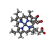
Mass: 670.104 Da / Num. of mol.: 4 / Source method: obtained synthetically / Formula: C36H36N4O5Zn / Feature type: SUBJECT OF INVESTIGATION
#3: Chemical ChemComp-PO4 / PHOSPHATE ION


Mass: 94.971 Da / Num. of mol.: 2 / Source method: obtained synthetically / Formula: PO4
#4: Chemical ChemComp-EDO / 1,2-ETHANEDIOL / ETHYLENE GLYCOL


Mass: 62.068 Da / Num. of mol.: 2 / Source method: obtained synthetically / Formula: C2H6O2
#5: Chemical ChemComp-PGE / TRIETHYLENE GLYCOL


Mass: 150.173 Da / Num. of mol.: 2 / Source method: obtained synthetically / Formula: C6H14O4
#6: Water ChemComp-HOH / water


Mass: 18.015 Da / Num. of mol.: 9 / Source method: isolated from a natural source / Formula: H2O

-
Details

Has ligand of interestY

-
Experimental details

-
Experiment

ExperimentMethod: X-RAY DIFFRACTION / Number of used crystals: 1

-
Sample preparation

CrystalDensity Matthews: 2.22 Å3/Da / Density % sol: 44.56 %
Crystal growTemperature: 298.15 K / Method: vapor diffusion, hanging drop / Details: 30% PEG 3350, 100mM Ammonium sulfate

-
Data collection

DiffractionMean temperature: 100 K / Serial crystal experiment: N
Diffraction sourceSource: SYNCHROTRON / Site: ALS / Beamline: 5.0.2 / Wavelength: 0.976 Å
DetectorType: DECTRIS PILATUS3 2M / Detector: PIXEL / Date: Sep 25, 2021
RadiationProtocol: SINGLE WAVELENGTH / Monochromatic (M) / Laue (L): M / Scattering type: x-ray
Radiation wavelengthWavelength: 0.976 Å / Relative weight: 1
ReflectionResolution: 2.07→49.26 Å / Num. obs: 52741 / % possible obs: 92.3 % / Redundancy: 3.5 % / Biso Wilson estimate: 52.92 Å2 / CC1/2: 0.998 / Rmerge(I) obs: 0.067 / Rpim(I) all: 0.043 / Rrim(I) all: 0.081 / Net I/σ(I): 7.5 / Num. measured all: 182543 / Scaling rejects: 2
Reflection shell

Diffraction-ID: 1 / Redundancy: 3.4 %

Resolution (Å)Rmerge(I) obsNum. measured allNum. unique obsCC1/2Rpim(I) allRrim(I) allNet I/σ(I) obs% possible all
2.07-2.131.297911027080.3980.8341.550.960.2
9.02-49.260.03522166520.9960.0230.04224.195.5

-
Processing

Software
NameVersionClassification
PHENIX1.2refinement
XDSdata reduction
Aimlessdata scaling
PDB_EXTRACT3.27data extraction
PHASERphasing
RefinementMethod to determine structure: MOLECULAR REPLACEMENT
Starting model: computationally generated

Resolution: 2.5→49.26 Å / SU ML: 0.31 / Cross valid method: THROUGHOUT / σ(F): 1.96 / Phase error: 29.22 / Stereochemistry target values: ML
RfactorNum. reflection% reflection
Rfree0.2519 1195 3.82 %
Rwork0.1988 30089 -
obs0.2009 31284 96.52 %
Solvent computationShrinkage radii: 0.9 Å / VDW probe radii: 1.11 Å / Solvent model: FLAT BULK SOLVENT MODEL
Displacement parametersBiso max: 142.1 Å2 / Biso mean: 64.1794 Å2 / Biso min: 33.34 Å2
Refinement stepCycle: final / Resolution: 2.5→49.26 Å
ProteinNucleic acidLigandSolventTotal
Num. atoms6684 0 206 9 6899
Biso mean--66.91 52.34 -
Num. residues----968
LS refinement shell

Refine-ID: X-RAY DIFFRACTION / Rfactor Rfree error: 0 / Total num. of bins used: 9

Resolution (Å)Rfactor RfreeNum. reflection RfreeRfactor RworkNum. reflection RworkNum. reflection all% reflection obs (%)
2.5-2.60.30911440.25463397354198
2.6-2.720.27821260.24363388351498
2.72-2.860.3081410.22763402354398
2.86-3.040.30761340.2373392352698
3.04-3.280.27561350.24313397353298
3.28-3.610.26241240.2033366349097
3.61-4.130.23841370.18043311344895
4.13-5.20.22091290.18113222335193
5.2-49.260.23031250.17643214333993
Refinement TLS params.Method: refined / Origin x: 0.5083 Å / Origin y: 5.2331 Å / Origin z: 27.7494 Å
111213212223313233
T0.2983 Å20.0036 Å2-0.0096 Å2-0.3654 Å20.0222 Å2--0.5071 Å2
L0.0772 °20.0197 °2-0.0443 °2-0.3297 °20.4392 °2--1.5589 °2
S0.0138 Å °-0.0559 Å °-0.0357 Å °0.0188 Å °-0.05 Å °-0.0108 Å °-0.0128 Å °-0.0366 Å °0.0428 Å °
Refinement TLS group
IDRefine-IDRefine TLS-IDSelection detailsAuth asym-IDAuth seq-ID
1X-RAY DIFFRACTION1allA1 - 244
2X-RAY DIFFRACTION1allC1 - 242
3X-RAY DIFFRACTION1allB0 - 244
4X-RAY DIFFRACTION1allD3 - 244
5X-RAY DIFFRACTION1allE11 - 28
6X-RAY DIFFRACTION1allF1 - 4
7X-RAY DIFFRACTION1allG1 - 2
8X-RAY DIFFRACTION1allH1 - 4

+
About Yorodumi

-
News

-
Feb 9, 2022. New format data for meta-information of EMDB entries

New format data for meta-information of EMDB entries

  • Version 3 of the EMDB header file is now the official format.
  • The previous official version 1.9 will be removed from the archive.

Related info.:EMDB header

External links:wwPDB to switch to version 3 of the EMDB data model

-
Aug 12, 2020. Covid-19 info

Covid-19 info

URL: https://pdbjlc1.pdbj.org/emnavi/covid19.php

New page: Covid-19 featured information page in EM Navigator.

Related info.:Covid-19 info / Mar 5, 2020. Novel coronavirus structure data

+
Mar 5, 2020. Novel coronavirus structure data

Novel coronavirus structure data

Related info.:Yorodumi Speices / Aug 12, 2020. Covid-19 info

External links:COVID-19 featured content - PDBj / Molecule of the Month (242):Coronavirus Proteases

+
Jan 31, 2019. EMDB accession codes are about to change! (news from PDBe EMDB page)

EMDB accession codes are about to change! (news from PDBe EMDB page)

  • The allocation of 4 digits for EMDB accession codes will soon come to an end. Whilst these codes will remain in use, new EMDB accession codes will include an additional digit and will expand incrementally as the available range of codes is exhausted. The current 4-digit format prefixed with “EMD-” (i.e. EMD-XXXX) will advance to a 5-digit format (i.e. EMD-XXXXX), and so on. It is currently estimated that the 4-digit codes will be depleted around Spring 2019, at which point the 5-digit format will come into force.
  • The EM Navigator/Yorodumi systems omit the EMD- prefix.

Related info.:Q: What is EMD? / ID/Accession-code notation in Yorodumi/EM Navigator

External links:EMDB Accession Codes are Changing Soon! / Contact to PDBj

+
Jul 12, 2017. Major update of PDB

Major update of PDB

  • wwPDB released updated PDB data conforming to the new PDBx/mmCIF dictionary.
  • This is a major update changing the version number from 4 to 5, and with Remediation, in which all the entries are updated.
  • In this update, many items about electron microscopy experimental information are reorganized (e.g. em_software).
  • Now, EM Navigator and Yorodumi are based on the updated data.

External links:wwPDB Remediation / Enriched Model Files Conforming to OneDep Data Standards Now Available in the PDB FTP Archive

-
Yorodumi

Thousand views of thousand structures

  • Yorodumi is a browser for structure data from EMDB, PDB, SASBDB, etc.
  • This page is also the successor to EM Navigator detail page, and also detail information page/front-end page for Omokage search.
  • The word "yorodu" (or yorozu) is an old Japanese word meaning "ten thousand". "mi" (miru) is to see.

Related info.:EMDB / PDB / SASBDB / Comparison of 3 databanks / Yorodumi Search / Aug 31, 2016. New EM Navigator & Yorodumi / Yorodumi Papers / Jmol/JSmol / Function and homology information / Changes in new EM Navigator and Yorodumi

Read more