 Movie
Movie Controller

[English] 日本語
 Yorodumi
- PDB-8gju: Crystal structure of human methylmalonyl-CoA mutase (MMUT) in com... -
+ Open data

- Basic information
| Entry | Database: PDB / ID: 8gju | |||||||||
|---|---|---|---|---|---|---|---|---|---|---|
| Title | Crystal structure of human methylmalonyl-CoA mutase (MMUT) in complex with methylmalonic acidemia type A protein (MMAA), coenzyme A, and GDP | |||||||||
 Components Components |
| |||||||||
 Keywords Keywords | Isomerase/Hydrolase / protein-protein complex / isomerase / GTPase / G-protein / cobalamin / vitamin B12 / transport / organometallic cofactor / Isomerase-Hydrolase complex | |||||||||
| Function / homology |  Function and homology information Function and homology informationDefective MMAA causes MMA, cblA type / Defective MUT causes MMAM / : / methylmalonyl-CoA mutase / methylmalonyl-CoA mutase activity / Cobalamin (Cbl) metabolism / cobalamin metabolic process / Propionyl-CoA catabolism / sulfur compound metabolic process / Hydrolases; Acting on acid anhydrides ...Defective MMAA causes MMA, cblA type / Defective MUT causes MMAM / : / methylmalonyl-CoA mutase / methylmalonyl-CoA mutase activity / Cobalamin (Cbl) metabolism / cobalamin metabolic process / Propionyl-CoA catabolism / sulfur compound metabolic process / Hydrolases; Acting on acid anhydrides / cobalamin binding / molecular carrier activity / mitochondrial matrix / GTPase activity / GTP binding / protein homodimerization activity / mitochondrion / metal ion binding / identical protein binding / cytoplasm / cytosol Similarity search - Function | |||||||||
| Biological species |  Homo sapiens (human) Homo sapiens (human) | |||||||||
| Method |  X-RAY DIFFRACTION / SYNCHROTRON / MOLECULAR REPLACEMENT / Resolution: 2.79 Å X-RAY DIFFRACTION / SYNCHROTRON / MOLECULAR REPLACEMENT / Resolution: 2.79 Å | |||||||||
 Authors Authors | Mascarenhas, R.M. / Ruetz, M. / Gouda, H. / Yaw, M. / Banerjee, R. | |||||||||
| Funding support |  United States, 2items United States, 2items
| |||||||||
 Citation Citation | Journal: Nat Commun / Year: 2023 Title: Architecture of the human G-protein-methylmalonyl-CoA mutase nanoassembly for B 12 delivery and repair. Authors: Mascarenhas, R. / Ruetz, M. / Gouda, H. / Heitman, N. / Yaw, M. / Banerjee, R. | |||||||||
| History |
|
- Structure visualization
| Structure viewer | Molecule: MolmilJmol/JSmol |
|---|
- Downloads & links
-Download
| PDBx/mmCIF format | 8gju.cif.gz | 771.1 KB | Display | PDBx/mmCIF format |
|---|---|---|---|---|
| PDB format | pdb8gju.ent.gz | 619.6 KB | Display | PDB format |
| PDBx/mmJSON format | 8gju.json.gz | Tree view | PDBx/mmJSON format | |
| Others |  Other downloads Other downloads |
-Validation report
| Arichive directory | https://data.pdbj.org/pub/pdb/validation_reports/gj/8gjuftp://data.pdbj.org/pub/pdb/validation_reports/gj/8gju | HTTPS FTP |
|---|
-Related structure data
| Related structure data | 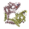 2wwwS S: Starting model for refinement |
|---|---|
| Similar structure data |
-Links
 PDBj
PDBj


- Assembly
| Deposited unit | 
| ||||||||
|---|---|---|---|---|---|---|---|---|---|
| 1 | 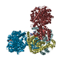
| ||||||||
| 2 | 
| ||||||||
| Unit cell |
|
-Components
-Protein , 2 types, 8 molecules DFABJKLH
| #1: Protein | Mass: 38718.867 Da / Num. of mol.: 4 Source method: isolated from a genetically manipulated source Source: (gene. exp.) Homo sapiens (human) / Gene: MMAA / Production host:  References: UniProt: Q8IVH4, Hydrolases; Acting on acid anhydrides #2: Protein | Mass: 83009.969 Da / Num. of mol.: 4 Source method: isolated from a genetically manipulated source Source: (gene. exp.) Homo sapiens (human) / Gene: MUT / Production host: |
|---|
-Non-polymers , 4 types, 25 molecules 


| #3: Chemical | ChemComp-GDP /  Type: RNA linking / Mass: 443.201 Da / Num. of mol.: 4 / Source method: obtained synthetically / Formula: C10H15N5O11P2 / Feature type: SUBJECT OF INVESTIGATION / Comment: GDP, energy-carrying molecule*YM Type: RNA linking / Mass: 443.201 Da / Num. of mol.: 4 / Source method: obtained synthetically / Formula: C10H15N5O11P2 / Feature type: SUBJECT OF INVESTIGATION / Comment: GDP, energy-carrying molecule*YM#4: Chemical | ChemComp-MG /  Mass: 24.305 Da / Num. of mol.: 4 / Source method: obtained synthetically / Formula: Mg Mass: 24.305 Da / Num. of mol.: 4 / Source method: obtained synthetically / Formula: Mg#5: Chemical | ChemComp-COA /  Mass: 767.534 Da / Num. of mol.: 4 / Source method: isolated from a natural source / Formula: C21H36N7O16P3S / Feature type: SUBJECT OF INVESTIGATION Mass: 767.534 Da / Num. of mol.: 4 / Source method: isolated from a natural source / Formula: C21H36N7O16P3S / Feature type: SUBJECT OF INVESTIGATION#6: Water | ChemComp-HOH / | Mass: 18.015 Da / Num. of mol.: 13 / Source method: isolated from a natural source / Formula: H2O |
|---|
-Details
| Has ligand of interest | Y |
|---|
-Experimental details
-Experiment
| Experiment | Method: X-RAY DIFFRACTION / Number of used crystals: 1 |
|---|
- Sample preparation
| Crystal | Density Matthews: 2.56 Å3/Da / Density % sol: 51.99 % |
|---|---|
| Crystal grow | Temperature: 293 K / Method: vapor diffusion, hanging drop Details: Morpheus1-F11 (Molecular Dimensions): 120 mM monosaccharides mix, 100 mM buffer system 3, pH 8.5, 30% precipitant mix 3 |
-Data collection
| Diffraction | Mean temperature: 80 K / Serial crystal experiment: N |
|---|---|
| Diffraction source | Source: SYNCHROTRON / Site: APS / Beamline: 23-ID-B / Wavelength: 1.033 Å |
| Detector | Type: DECTRIS EIGER X 16M / Detector: PIXEL / Date: Nov 2, 2021 |
| Radiation | Protocol: SINGLE WAVELENGTH / Monochromatic (M) / Laue (L): M / Scattering type: x-ray |
| Radiation wavelength | Wavelength: 1.033 Å / Relative weight: 1 |
| Reflection | Resolution: 2.7→117 Å / Num. obs: 74830 / % possible obs: 89 % / Redundancy: 3.1 % / CC1/2: 0.991 / Rmerge(I) obs: 0.137 / Rpim(I) all: 0.092 / Net I/σ(I): 5.2 |
| Reflection shell | Resolution: 2.794→3.074 Å / Rmerge(I) obs: 0.775 / Num. unique obs: 2737 / CC1/2: 0.562 / Rpim(I) all: 0.504 |
- Processing
| Software |
| ||||||||||||||||||||||||||||||||||||||||||||||||||||||||||||||||||||||||||||||||||||||||||||||||||||||||||||||||||||||||||||||||||||||||||||||||||||||||||||||||||||||||||||||||||||||||||||||||||||
|---|---|---|---|---|---|---|---|---|---|---|---|---|---|---|---|---|---|---|---|---|---|---|---|---|---|---|---|---|---|---|---|---|---|---|---|---|---|---|---|---|---|---|---|---|---|---|---|---|---|---|---|---|---|---|---|---|---|---|---|---|---|---|---|---|---|---|---|---|---|---|---|---|---|---|---|---|---|---|---|---|---|---|---|---|---|---|---|---|---|---|---|---|---|---|---|---|---|---|---|---|---|---|---|---|---|---|---|---|---|---|---|---|---|---|---|---|---|---|---|---|---|---|---|---|---|---|---|---|---|---|---|---|---|---|---|---|---|---|---|---|---|---|---|---|---|---|---|---|---|---|---|---|---|---|---|---|---|---|---|---|---|---|---|---|---|---|---|---|---|---|---|---|---|---|---|---|---|---|---|---|---|---|---|---|---|---|---|---|---|---|---|---|---|---|---|---|---|
| Refinement | Method to determine structure: MOLECULAR REPLACEMENT Starting model: 2WWW Resolution: 2.79→80.62 Å / SU ML: 0.31 / Cross valid method: THROUGHOUT / σ(F): 1.36 / Phase error: 33.92 / Stereochemistry target values: ML
| ||||||||||||||||||||||||||||||||||||||||||||||||||||||||||||||||||||||||||||||||||||||||||||||||||||||||||||||||||||||||||||||||||||||||||||||||||||||||||||||||||||||||||||||||||||||||||||||||||||
| Solvent computation | Shrinkage radii: 0.9 Å / VDW probe radii: 1.1 Å / Solvent model: FLAT BULK SOLVENT MODEL | ||||||||||||||||||||||||||||||||||||||||||||||||||||||||||||||||||||||||||||||||||||||||||||||||||||||||||||||||||||||||||||||||||||||||||||||||||||||||||||||||||||||||||||||||||||||||||||||||||||
| Refinement step | Cycle: LAST / Resolution: 2.79→80.62 Å
| ||||||||||||||||||||||||||||||||||||||||||||||||||||||||||||||||||||||||||||||||||||||||||||||||||||||||||||||||||||||||||||||||||||||||||||||||||||||||||||||||||||||||||||||||||||||||||||||||||||
| Refine LS restraints |
| ||||||||||||||||||||||||||||||||||||||||||||||||||||||||||||||||||||||||||||||||||||||||||||||||||||||||||||||||||||||||||||||||||||||||||||||||||||||||||||||||||||||||||||||||||||||||||||||||||||
| LS refinement shell |
|
