 Movie
Movie Controller

[English] 日本語
 Yorodumi
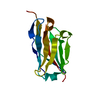
- PDB-8gjr: Crystal Structure of Nanobody VHH114 Bound to Its Antigen PA14 Cif -
+ Open data

- Basic information
| Entry | Database: PDB / ID: 8gjr | ||||||||||||||||||
|---|---|---|---|---|---|---|---|---|---|---|---|---|---|---|---|---|---|---|---|
| Title | Crystal Structure of Nanobody VHH114 Bound to Its Antigen PA14 Cif | ||||||||||||||||||
 Components Components |
| ||||||||||||||||||
 Keywords Keywords | IMMUNE SYSTEM / Pseudomonas aeruginosa / nanobody VHH immunoglobulin domain / CFTR inhibitory factor (Cif) | ||||||||||||||||||
| Function / homology | Alpha/beta hydrolase family / Alpha/beta hydrolase fold-1 / Alpha/Beta hydrolase fold / CITRATE ANION / CFTR inhibitory factor Function and homology information Function and homology information | ||||||||||||||||||
| Biological species |  Pseudomonas aeruginosa PA14 (bacteria) Pseudomonas aeruginosa PA14 (bacteria) | ||||||||||||||||||
| Method |  X-RAY DIFFRACTION / SYNCHROTRON / MOLECULAR REPLACEMENT / Resolution: 1.85 Å X-RAY DIFFRACTION / SYNCHROTRON / MOLECULAR REPLACEMENT / Resolution: 1.85 Å | ||||||||||||||||||
 Authors Authors | Simard, A.R. / Madden, D.R. | ||||||||||||||||||
| Funding support |  United States, 5items United States, 5items
| ||||||||||||||||||
 Citation Citation | Journal: To Be Published Title: Crystal Structure of Nanobody VHH114 Bound to Its Antigen PA14 Cif Authors: Simard, A.R. / Madden, D.R. #1: Journal: Anal Chim Acta / Year: 2019 Title: Nanobody-based binding assay for the discovery of potent inhibitors of CFTR inhibitory factor (Cif). Authors: Vasylieva, N. / Kitamura, S. / Dong, J. / Barnych, B. / Hvorecny, K.L. / Madden, D.R. / Gee, S.J. / Wolan, D.W. / Morisseau, C. / Hammock, B.D. #2: Journal: Acta Crystallogr D Biol Crystallogr / Year: 2012 Title: Towards automated crystallographic structure refinement with phenix.refine. Authors: Afonine, P.V. / Grosse-Kunstleve, R.W. / Echols, N. / Headd, J.J. / Moriarty, N.W. / Mustyakimov, M. / Terwilliger, T.C. / Urzhumtsev, A. / Zwart, P.H. / Adams, P.D. | ||||||||||||||||||
| History |
|
- Structure visualization
| Structure viewer | Molecule: MolmilJmol/JSmol |
|---|
- Downloads & links
-Download
| PDBx/mmCIF format | 8gjr.cif.gz | 114.4 KB | Display | PDBx/mmCIF format |
|---|---|---|---|---|
| PDB format | pdb8gjr.ent.gz | 78.1 KB | Display | PDB format |
| PDBx/mmJSON format | 8gjr.json.gz | Tree view | PDBx/mmJSON format | |
| Others |  Other downloads Other downloads |
-Validation report
| Arichive directory | https://data.pdbj.org/pub/pdb/validation_reports/gj/8gjrftp://data.pdbj.org/pub/pdb/validation_reports/gj/8gjr | HTTPS FTP |
|---|
-Related structure data
| Related structure data | |
|---|---|
| Similar structure data |


-Links
 PDBj
PDBj


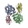
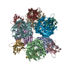
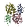
- Assembly
| Deposited unit | 
| |||||||||||||||||||||
|---|---|---|---|---|---|---|---|---|---|---|---|---|---|---|---|---|---|---|---|---|---|---|
| 1 | 
| |||||||||||||||||||||
| Unit cell |
| |||||||||||||||||||||
| Components on special symmetry positions |
|
-Components
| #1: Protein | Mass: 33335.828 Da / Num. of mol.: 1 Source method: isolated from a genetically manipulated source Source: (gene. exp.) Pseudomonas aeruginosa PA14 (bacteria) / Gene: PA14_26090 / Plasmid: pET16b / Details (production host): N-terminal 10XHis-SUMO fusion / Production host: | ||||||
|---|---|---|---|---|---|---|---|
| #2: Antibody | Mass: 13525.262 Da / Num. of mol.: 1 Source method: isolated from a genetically manipulated source Source: (gene. exp.) | ||||||
| #3: Chemical |   Mass: 189.100 Da / Num. of mol.: 2 / Source method: obtained synthetically / Formula: C6H5O7 Mass: 189.100 Da / Num. of mol.: 2 / Source method: obtained synthetically / Formula: C6H5O7#4: Water | ChemComp-HOH / |  Mass: 18.015 Da / Num. of mol.: 335 / Source method: isolated from a natural source / Formula: H2O Mass: 18.015 Da / Num. of mol.: 335 / Source method: isolated from a natural source / Formula: H2OHas ligand of interest | N | Has protein modification | Y | |
-Experimental details
-Experiment
| Experiment | Method: X-RAY DIFFRACTION / Number of used crystals: 1 |
|---|
- Sample preparation
| Crystal | Density Matthews: 3.09 Å3/Da / Density % sol: 60.21 % / Description: hexagonal rods |
|---|---|
| Crystal grow | Temperature: 292.8 K / Method: vapor diffusion, hanging drop / pH: 3.5 Details: 13.5 % (w/v) PEG6K, 2.5 % (v/v) ethylene glycol, 100 mM sodium citrate pH 3.5 |
-Data collection
| Diffraction | Mean temperature: 100 K / Serial crystal experiment: N |
|---|---|
| Diffraction source | Source: SYNCHROTRON / Site: NSLS-II / Beamline: 17-ID-1 / Wavelength: 0.920112 Å |
| Detector | Type: DECTRIS EIGER X 9M / Detector: PIXEL / Date: Feb 12, 2023 |
| Radiation | Protocol: SINGLE WAVELENGTH / Monochromatic (M) / Laue (L): M / Scattering type: x-ray |
| Radiation wavelength | Wavelength: 0.920112 Å / Relative weight: 1 |
| Reflection | Resolution: 1.85→27.62 Å / Num. obs: 50643 / % possible obs: 99.91 % / Redundancy: 20.2 % / Biso Wilson estimate: 32 Å2 / CC1/2: 0.999 / CC star: 1 / Rmerge(I) obs: 0.139 / Rpim(I) all: 0.03181 / Rrim(I) all: 0.1427 / Net I/σ(I): 16.31 |
| Reflection shell | Resolution: 1.85→1.916 Å / Redundancy: 20.8 % / Rmerge(I) obs: 2.404 / Mean I/σ(I) obs: 1.23 / Num. unique obs: 5007 / CC1/2: 0.618 / CC star: 0.874 / Rpim(I) all: 0.539 / Rrim(I) all: 2.465 / % possible all: 99.96 |
- Processing
| Software |
| |||||||||||||||||||||||||||||||||||||||||||||||||||||||||||||||||||||||||||||||||||||||||||||||||||||||||||||||||||||||||||||||||||||
|---|---|---|---|---|---|---|---|---|---|---|---|---|---|---|---|---|---|---|---|---|---|---|---|---|---|---|---|---|---|---|---|---|---|---|---|---|---|---|---|---|---|---|---|---|---|---|---|---|---|---|---|---|---|---|---|---|---|---|---|---|---|---|---|---|---|---|---|---|---|---|---|---|---|---|---|---|---|---|---|---|---|---|---|---|---|---|---|---|---|---|---|---|---|---|---|---|---|---|---|---|---|---|---|---|---|---|---|---|---|---|---|---|---|---|---|---|---|---|---|---|---|---|---|---|---|---|---|---|---|---|---|---|---|---|
| Refinement | Method to determine structure: MOLECULAR REPLACEMENT / Resolution: 1.85→27.62 Å / SU ML: 0.2244 / Cross valid method: FREE R-VALUE / σ(F): 1.36 / Phase error: 20.157 Stereochemistry target values: GeoStd + Monomer Library + CDL v1.2 Details: Atoms modeled with zero occupancy could not be placed with confidence and were selected for zero-occupancy flagging after manual inspection of the 2Fo-Fc map at a 0.5-sigma cutoff.
| |||||||||||||||||||||||||||||||||||||||||||||||||||||||||||||||||||||||||||||||||||||||||||||||||||||||||||||||||||||||||||||||||||||
| Solvent computation | Shrinkage radii: 0.9 Å / VDW probe radii: 1.1 Å / Solvent model: FLAT BULK SOLVENT MODEL | |||||||||||||||||||||||||||||||||||||||||||||||||||||||||||||||||||||||||||||||||||||||||||||||||||||||||||||||||||||||||||||||||||||
| Displacement parameters | Biso mean: 33.57 Å2 | |||||||||||||||||||||||||||||||||||||||||||||||||||||||||||||||||||||||||||||||||||||||||||||||||||||||||||||||||||||||||||||||||||||
| Refinement step | Cycle: LAST / Resolution: 1.85→27.62 Å
| |||||||||||||||||||||||||||||||||||||||||||||||||||||||||||||||||||||||||||||||||||||||||||||||||||||||||||||||||||||||||||||||||||||
| Refine LS restraints |
| |||||||||||||||||||||||||||||||||||||||||||||||||||||||||||||||||||||||||||||||||||||||||||||||||||||||||||||||||||||||||||||||||||||
| LS refinement shell |
|
