 Movie
Movie Controller


+ Open data

- Basic information
| Entry | Database: PDB / ID: 8e1c | ||||||||||||||||||
|---|---|---|---|---|---|---|---|---|---|---|---|---|---|---|---|---|---|---|---|
| Title | Crystal Structure of Nanobody VHH222 Specific for PA14 Cif | ||||||||||||||||||
 Components Components | Nanobody VHH222 | ||||||||||||||||||
 Keywords Keywords | IMMUNE SYSTEM / Pseudomonas aeruginosa / nanobody VHH / immunoglobulin domain / CFTR inhibitory factor (Cif) | ||||||||||||||||||
| Biological species |  | ||||||||||||||||||
| Method |  X-RAY DIFFRACTION / SYNCHROTRON / MOLECULAR REPLACEMENT / Resolution: 1.9 Å X-RAY DIFFRACTION / SYNCHROTRON / MOLECULAR REPLACEMENT / Resolution: 1.9 Å | ||||||||||||||||||
 Authors Authors | Simard, A.R. / Taher, N.M. / Balsam, S.S. / Madden, D.R. | ||||||||||||||||||
| Funding support |  United States, 5items United States, 5items
| ||||||||||||||||||
 Citation Citation | Journal: To Be Published Title: Crystal Structure of Nanobody VHH222 Specific for PA14 Cif Authors: Simard, A.R. / Madden, D.R. #1: Journal: Anal Chim Acta / Year: 2019 Title: Nanobody-based binding assay for the discovery of potent inhibitors of CFTR inhibitory factor (Cif). Authors: Vasylieva, N. / Kitamura, S. / Dong, J. / Barnych, B. / Hvorecny, K.L. / Madden, D.R. / Gee, S.J. / Wolan, D.W. / Morisseau, C. / Hammock, B.D. | ||||||||||||||||||
| History |
|
- Structure visualization
| Structure viewer | Molecule:  MolmilJmol/JSmol MolmilJmol/JSmol |
|---|
- Downloads & links
-Download
| PDBx/mmCIF format | 8e1c.cif.gz | 38.5 KB | Display | PDBx/mmCIF format |
|---|---|---|---|---|
| PDB format | pdb8e1c.ent.gz | 23.9 KB | Display | PDB format |
| PDBx/mmJSON format | 8e1c.json.gz | Tree view | PDBx/mmJSON format | |
| Others |  Other downloads Other downloads |
-Validation report
| Arichive directory | https://data.pdbj.org/pub/pdb/validation_reports/e1/8e1cftp://data.pdbj.org/pub/pdb/validation_reports/e1/8e1c | HTTPS FTP |
|---|
-Related structure data
| Related structure data | 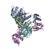 3ezjS S: Starting model for refinement |
|---|
-Links
 PDBj
PDBj

- Assembly
| Deposited unit | 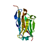
| ||||||||||||
|---|---|---|---|---|---|---|---|---|---|---|---|---|---|
| 1 |
| ||||||||||||
| Unit cell |
| ||||||||||||
| Components on special symmetry positions |
|
-Components
| #1: Antibody | Mass: 13531.143 Da / Num. of mol.: 1 Source method: isolated from a genetically manipulated source Source: (gene. exp.)  |
|---|---|
| #2: Water | ChemComp-HOH /  Mass: 18.015 Da / Num. of mol.: 57 / Source method: isolated from a natural source / Formula: H2O Mass: 18.015 Da / Num. of mol.: 57 / Source method: isolated from a natural source / Formula: H2O |
| Has protein modification | Y |
-Experimental details
-Experiment
| Experiment | Method: X-RAY DIFFRACTION / Number of used crystals: 1 |
|---|
- Sample preparation
| Crystal | Density Matthews: 2.73 Å3/Da / Density % sol: 54.87 % |
|---|---|
| Crystal grow | Temperature: 292.8 K / Method: vapor diffusion, sitting drop / pH: 6 Details: 100 mM succinic acid/sodium phosphate monobasic/glycine 2:7:7 (SPG) pH 6.0, 25% (w/v) PEG1500 |
-Data collection
| Diffraction | Mean temperature: 100 K / Serial crystal experiment: N |
|---|---|
| Diffraction source | Source: SYNCHROTRON / Site: NSLS-II / Beamline: 17-ID-2 / Wavelength: 0.978636 Å |
| Detector | Type: DECTRIS EIGER X 16M / Detector: PIXEL / Date: Jul 29, 2020 |
| Radiation | Protocol: SINGLE WAVELENGTH / Monochromatic (M) / Laue (L): M / Scattering type: x-ray |
| Radiation wavelength | Wavelength: 0.978636 Å / Relative weight: 1 |
| Reflection | Resolution: 1.9→41.9 Å / Num. obs: 12038 / % possible obs: 99.48 % / Redundancy: 9.5 % / Biso Wilson estimate: 35.72 Å2 / CC1/2: 0.999 / CC star: 1 / Rmerge(I) obs: 0.07468 / Rpim(I) all: 0.02545 / Rrim(I) all: 0.07902 / Net I/σ(I): 16.76 |
| Reflection shell | Resolution: 1.9→1.968 Å / Redundancy: 7.6 % / Rmerge(I) obs: 1.286 / Mean I/σ(I) obs: 1.58 / Num. unique obs: 1142 / CC1/2: 0.838 / CC star: 0.955 / Rpim(I) all: 0.4838 / Rrim(I) all: 1.379 / % possible all: 97.35 |
- Processing
| Software |
| |||||||||||||||||||||||||||||||||||
|---|---|---|---|---|---|---|---|---|---|---|---|---|---|---|---|---|---|---|---|---|---|---|---|---|---|---|---|---|---|---|---|---|---|---|---|---|
| Refinement | Method to determine structure: MOLECULAR REPLACEMENT Starting model: 3EZJ Resolution: 1.9→41.9 Å / SU ML: 0.2361 / Cross valid method: FREE R-VALUE / σ(F): 1.37 / Phase error: 30.2802 Stereochemistry target values: GeoStd + Monomer Library + CDL v1.2 Details: Positive electron-density peaks are observed where residues 104:106 of Chain A would be located, but the placement of main-chain atoms could not be resolved.
| |||||||||||||||||||||||||||||||||||
| Solvent computation | Shrinkage radii: 0.9 Å / VDW probe radii: 1.1 Å / Solvent model: FLAT BULK SOLVENT MODEL | |||||||||||||||||||||||||||||||||||
| Displacement parameters | Biso mean: 42.92 Å2 | |||||||||||||||||||||||||||||||||||
| Refinement step | Cycle: LAST / Resolution: 1.9→41.9 Å
| |||||||||||||||||||||||||||||||||||
| Refine LS restraints |
| |||||||||||||||||||||||||||||||||||
| LS refinement shell |
|
