| 登録情報 | データベース: PDB / ID: 8gji
|
|---|
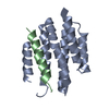
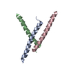
| タイトル | De novo design of high-affinity protein binders to bioactive helical peptides |
|---|
 要素 要素 | |
|---|
 キーワード キーワード | DE NOVO PROTEIN / Alpha-helical peptides / protein design / diffusion / deep learning |
|---|
| 機能・相同性 |  機能・相同性情報 機能・相同性情報
glucagon receptor binding / regulation of insulin secretion / positive regulation of insulin secretion involved in cellular response to glucose stimulus / response to activity / hormone activity / adenylate cyclase-modulating G protein-coupled receptor signaling pathway / glucose homeostasis / negative regulation of apoptotic process / : 類似検索 - 分子機能 Glucagon / Glucagon/GIP/secretin/VIP / Peptide hormone / Glucagon / GIP / secretin / VIP family signature. / Glucagon like hormones類似検索 - ドメイン・相同性 |
|---|
| 生物種 | synthetic construct (人工物) |
|---|
| 手法 |  X線回折 / シンクロトロン / 分子置換 / 解像度: 1.81 Å X線回折 / シンクロトロン / 分子置換 / 解像度: 1.81 Å |
|---|
 データ登録者 データ登録者 | Torres, S.V. / Leung, P.J.Y. / Bera, A.K. / Baker, D. / Kang, A. |
|---|
| 資金援助 |  米国, 2件 米国, 2件 | 組織 | 認可番号 | 国 |
|---|
| National Science Foundation (NSF, United States) | | 米国 | | Howard Hughes Medical Institute (HHMI) | | 米国 |
|
|---|
 引用 引用 | ジャーナル: Nature / 年: 2024
タイトル: De novo design of high-affinity binders of bioactive helical peptides.
著者: Vazquez Torres, S. / Leung, P.J.Y. / Venkatesh, P. / Lutz, I.D. / Hink, F. / Huynh, H.H. / Becker, J. / Yeh, A.H. / Juergens, D. / Bennett, N.R. / Hoofnagle, A.N. / Huang, E. / MacCoss, M.J. ...著者: Vazquez Torres, S. / Leung, P.J.Y. / Venkatesh, P. / Lutz, I.D. / Hink, F. / Huynh, H.H. / Becker, J. / Yeh, A.H. / Juergens, D. / Bennett, N.R. / Hoofnagle, A.N. / Huang, E. / MacCoss, M.J. / Exposit, M. / Lee, G.R. / Bera, A.K. / Kang, A. / De La Cruz, J. / Levine, P.M. / Li, X. / Lamb, M. / Gerben, S.R. / Murray, A. / Heine, P. / Korkmaz, E.N. / Nivala, J. / Stewart, L. / Watson, J.L. / Rogers, J.M. / Baker, D. |
|---|
| 履歴 | | 登録 | 2023年3月15日 | 登録サイト: RCSB / 処理サイト: RCSB |
|---|
| 改定 1.0 | 2024年1月10日 | Provider: repository / タイプ: Initial release |
|---|
| 改定 1.1 | 2024年2月14日 | Group: Database references / カテゴリ: citation / citation_author
Item: _citation.journal_volume / _citation.page_first ..._citation.journal_volume / _citation.page_first / _citation.page_last / _citation.year / _citation_author.name |
|---|
|
|---|
 ムービー
ムービー コントローラー


 データを開く
 基本情報
基本情報 構造の表示
構造の表示 ダウンロードとリンク
 その他のダウンロード
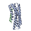
 PDBj
PDBj


 集合体
集合体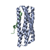

 分子量: 18.015 Da / 分子数: 24 / 由来タイプ: 天然 / 式: H2O
 試料調製
試料調製 解析
解析