| 登録情報 | データベース: PDB / ID: 8gdw
|
|---|
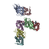
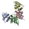
| タイトル | Crystal structure of Domain Related to Iron (DRI) from cyanobacteria |
|---|
 要素 要素 | Ssr1698 protein |
|---|
 キーワード キーワード | METAL BINDING PROTEIN / Heme binding protein / DUF2470 / heme homeostasis / succinate dehydrogenase / zinc and heme binding sites |
|---|
| 機能・相同性 | Domain of unknown function DUF2470 / Domain of unknown function (DUF2470) / Haem oxygenase HugZ-like superfamily / metal ion binding / Ssr1698 protein 機能・相同性情報 機能・相同性情報 |
|---|
| 生物種 |   Synechocystis sp. PCC 6803 (バクテリア) Synechocystis sp. PCC 6803 (バクテリア) |
|---|
| 手法 |  X線回折 / シンクロトロン / 分子置換 / 解像度: 2.35 Å X線回折 / シンクロトロン / 分子置換 / 解像度: 2.35 Å |
|---|
 データ登録者 データ登録者 | Kumaran, D. / Grosjean, N. / Blaby, E.C. |
|---|
| 資金援助 |  米国, 1件 米国, 1件 | 組織 | 認可番号 | 国 |
|---|
| Department of Energy (DOE, United States) | Quantitative Plant Science Initiative SFA | 米国 |
|
|---|
 引用 引用 | ジャーナル: Nat Commun / 年: 2024
タイトル: A hemoprotein with a zinc-mirror heme site ties heme availability to carbon metabolism in cyanobacteria.
著者: Grosjean, N. / Yee, E.F. / Kumaran, D. / Chopra, K. / Abernathy, M. / Biswas, S. / Byrnes, J. / Kreitler, D.F. / Cheng, J.F. / Ghosh, A. / Almo, S.C. / Iwai, M. / Niyogi, K.K. / Pakrasi, H.B. ...著者: Grosjean, N. / Yee, E.F. / Kumaran, D. / Chopra, K. / Abernathy, M. / Biswas, S. / Byrnes, J. / Kreitler, D.F. / Cheng, J.F. / Ghosh, A. / Almo, S.C. / Iwai, M. / Niyogi, K.K. / Pakrasi, H.B. / Sarangi, R. / van Dam, H. / Yang, L. / Blaby, I.K. / Blaby-Haas, C.E. |
|---|
| 履歴 | | 登録 | 2023年3月6日 | 登録サイト: RCSB / 処理サイト: RCSB |
|---|
| 改定 1.0 | 2024年3月13日 | Provider: repository / タイプ: Initial release |
|---|
| 改定 1.1 | 2024年4月24日 | Group: Database references / カテゴリ: citation / citation_author
Item: _citation.country / _citation.journal_abbrev ..._citation.country / _citation.journal_abbrev / _citation.journal_id_CSD / _citation.journal_id_ISSN / _citation.journal_volume / _citation.page_first / _citation.page_last / _citation.pdbx_database_id_DOI / _citation.pdbx_database_id_PubMed / _citation.title / _citation.year |
|---|
|
|---|
 ムービー
ムービー コントローラー


 データを開く
 基本情報
基本情報 構造の表示
構造の表示 ダウンロードとリンク
 その他のダウンロード
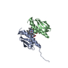

 PDBj
PDBj 集合体
集合体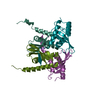

 分子量: 65.409 Da / 分子数: 4 / 由来タイプ: 合成 / 式: Zn / タイプ: SUBJECT OF INVESTIGATION
 分子量: 18.015 Da / 分子数: 34 / 由来タイプ: 天然 / 式: H2O
 試料調製
試料調製 解析
解析