 Movie
Movie Controller


+ Open data

- Basic information
| Entry | Database: PDB / ID: 8fm6 | ||||||
|---|---|---|---|---|---|---|---|
| Title | Dri1 hemoprotein variant H21A with a zinc-mirror heme site | ||||||
 Components Components | Ssr1698 protein | ||||||
 Keywords Keywords | METAL BINDING PROTEIN / heme / DRI domain | ||||||
| Function / homology | Domain of unknown function DUF2470 / Domain of unknown function (DUF2470) / Haem oxygenase HugZ-like superfamily / metal ion binding / HEME B/C / Ssr1698 protein Function and homology information Function and homology information | ||||||
| Biological species |  | ||||||
| Method |  X-RAY DIFFRACTION / SYNCHROTRON / MOLECULAR REPLACEMENT / Resolution: 2.85 Å X-RAY DIFFRACTION / SYNCHROTRON / MOLECULAR REPLACEMENT / Resolution: 2.85 Å | ||||||
 Authors Authors | Yee, E.F. / Blaby-Haas, C. | ||||||
| Funding support | 1items
| ||||||
 Citation Citation | Journal: Nat Commun / Year: 2024 Title: A hemoprotein with a zinc-mirror heme site ties heme availability to carbon metabolism in cyanobacteria. Authors: Grosjean, N. / Yee, E.F. / Kumaran, D. / Chopra, K. / Abernathy, M. / Biswas, S. / Byrnes, J. / Kreitler, D.F. / Cheng, J.F. / Ghosh, A. / Almo, S.C. / Iwai, M. / Niyogi, K.K. / Pakrasi, H.B. ...Authors: Grosjean, N. / Yee, E.F. / Kumaran, D. / Chopra, K. / Abernathy, M. / Biswas, S. / Byrnes, J. / Kreitler, D.F. / Cheng, J.F. / Ghosh, A. / Almo, S.C. / Iwai, M. / Niyogi, K.K. / Pakrasi, H.B. / Sarangi, R. / van Dam, H. / Yang, L. / Blaby, I.K. / Blaby-Haas, C.E. | ||||||
| History |
|
- Structure visualization
| Structure viewer | Molecule: MolmilJmol/JSmol |
|---|
- Downloads & links
-Download
| PDBx/mmCIF format | 8fm6.cif.gz | 102.2 KB | Display | PDBx/mmCIF format |
|---|---|---|---|---|
| PDB format | pdb8fm6.ent.gz | 69.9 KB | Display | PDB format |
| PDBx/mmJSON format | 8fm6.json.gz | Tree view | PDBx/mmJSON format | |
| Others |  Other downloads Other downloads |
-Validation report
| Arichive directory | https://data.pdbj.org/pub/pdb/validation_reports/fm/8fm6ftp://data.pdbj.org/pub/pdb/validation_reports/fm/8fm6 | HTTPS FTP |
|---|
-Related structure data
| Related structure data | 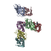 8gbkC  8gdwC 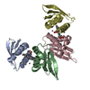 8gf4C C: citing same article ( |
|---|---|
| Similar structure data | |
| Other databases |
|
-Links
 PDBj
PDBj
- Assembly
| Deposited unit | 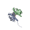
| ||||||||||||||||||||||||||||||
|---|---|---|---|---|---|---|---|---|---|---|---|---|---|---|---|---|---|---|---|---|---|---|---|---|---|---|---|---|---|---|---|
| 1 |
| ||||||||||||||||||||||||||||||
| Unit cell |
| ||||||||||||||||||||||||||||||
| Noncrystallographic symmetry (NCS) | NCS domain:
NCS domain segments:
|
-Components
| #1: Protein | Mass: 11239.646 Da / Num. of mol.: 2 / Mutation: H21A Source method: isolated from a genetically manipulated source Source: (gene. exp.) #2: Chemical | ChemComp-HEB / |   Mass: 618.503 Da / Num. of mol.: 1 / Source method: obtained synthetically / Formula: C34H34FeN4O4 / Feature type: SUBJECT OF INVESTIGATION Mass: 618.503 Da / Num. of mol.: 1 / Source method: obtained synthetically / Formula: C34H34FeN4O4 / Feature type: SUBJECT OF INVESTIGATIONHas ligand of interest | Y | |
|---|
-Experimental details
-Experiment
| Experiment | Method: X-RAY DIFFRACTION / Number of used crystals: 1 |
|---|
- Sample preparation
| Crystal | Density Matthews: 3.78 Å3/Da / Density % sol: 67.45 % |
|---|---|
| Crystal grow | Temperature: 298 K / Method: vapor diffusion, hanging drop / pH: 5 Details: 0.1 M sodium citrate, 0.1 M potassium phosphate, 34% PEG8000 |
-Data collection
| Diffraction | Mean temperature: 100 K / Serial crystal experiment: N |
|---|---|
| Diffraction source | Source: SYNCHROTRON / Site: NSLS-II  / Beamline: 17-ID-1 / Wavelength: 0.92 Å / Beamline: 17-ID-1 / Wavelength: 0.92 Å |
| Detector | Type: DECTRIS EIGER X 9M / Detector: PIXEL / Date: Oct 16, 2022 / Details: KB bimorph mirrors |
| Radiation | Monochromator: Si(111) DCM / Protocol: SINGLE WAVELENGTH / Monochromatic (M) / Laue (L): M / Scattering type: x-ray |
| Radiation wavelength | Wavelength: 0.92 Å / Relative weight: 1 |
| Reflection | Resolution: 2.85→36.76 Å / Num. obs: 8771 / % possible obs: 81.64 % / Redundancy: 10 % / Biso Wilson estimate: 81.6 Å2 / CC1/2: 0.998 / CC star: 1 / Rmerge(I) obs: 0.148 / Rpim(I) all: 0.049 / Rrim(I) all: 0.156 / Net I/σ(I): 10.7 |
| Reflection shell | Resolution: 2.851→3.05 Å / Redundancy: 12.8 % / Rmerge(I) obs: 2.395 / Mean I/σ(I) obs: 1.2 / Num. unique obs: 106 / CC1/2: 0.436 / Rpim(I) all: 0.691 / Rrim(I) all: 2.495 / % possible all: 63.7 |
- Processing
| Software |
| ||||||||||||||||||||||||||||||||||||||||
|---|---|---|---|---|---|---|---|---|---|---|---|---|---|---|---|---|---|---|---|---|---|---|---|---|---|---|---|---|---|---|---|---|---|---|---|---|---|---|---|---|---|
| Refinement | Method to determine structure: MOLECULAR REPLACEMENT / Resolution: 2.85→36.76 Å / SU ML: 0.3329 / Cross valid method: FREE R-VALUE / σ(F): 1.35 / Phase error: 28.9282 Stereochemistry target values: GeoStd + Monomer Library + CDL v1.2
| ||||||||||||||||||||||||||||||||||||||||
| Solvent computation | Shrinkage radii: 0.9 Å / VDW probe radii: 1.11 Å / Solvent model: FLAT BULK SOLVENT MODEL | ||||||||||||||||||||||||||||||||||||||||
| Displacement parameters | Biso mean: 77.57 Å2 | ||||||||||||||||||||||||||||||||||||||||
| Refinement step | Cycle: LAST / Resolution: 2.85→36.76 Å
| ||||||||||||||||||||||||||||||||||||||||
| Refine LS restraints |
| ||||||||||||||||||||||||||||||||||||||||
| Refine LS restraints NCS | Type: Torsion NCS / Rms dev position: 0.921678426611 Å | ||||||||||||||||||||||||||||||||||||||||
| LS refinement shell |
| ||||||||||||||||||||||||||||||||||||||||
| Refinement TLS params. | Method: refined / Origin x: -26.9976013583 Å / Origin y: 19.4440202137 Å / Origin z: -1.77762991529 Å
| ||||||||||||||||||||||||||||||||||||||||
| Refinement TLS group | Selection details: all |
