Entry Database : PDB / ID : 8g9dTitle Diphosphoinositol polyphosphate phosphohydrolase 1 (DIPP1/NUDT3) in complex with 5- phosphonodifluoroacetamide inositol pentakisphosphate (5-PCF2Am-InsP5), an analogue of 5-InsP7 Diphosphoinositol polyphosphate phosphohydrolase 1 Keywords / / / / / / Function / homology Function Domain/homology Component
/ / / / / / / / / / / / / / / / / / / / / / / / / / / / / / / / / / / / / / / / / / / / / Biological species Homo sapiens (human)Method / / / Resolution : 1.6 Å Authors Zong, G. / Wang, H. / Shears, S. Funding support Organization Grant number Country National Institutes of Health/National Institute of Environmental Health Sciences (NIH/NIEHS) 1ZIAES080046-31
Journal : Chemistry / Year : 2023Title : Fluorination Influences the Bioisostery of Myo-Inositol Pyrophosphate Analogs.Authors : Hostachy, S. / Wang, H. / Zong, G. / Franke, K. / Riley, A.M. / Schmieder, P. / Potter, B.V.L. / Shears, S.B. / Fiedler, D. History Deposition Feb 21, 2023 Deposition site / Processing site Revision 1.0 Jan 3, 2024 Provider / Type
 Movie
Movie Controller

 Yorodumi
 Open data
 Basic information
 Components
 Keywords
 Function and homology information
 Homo sapiens (human)
 X-RAY DIFFRACTION /
  Authors
 United States, 1items
  Citation
 Structure visualization
 Downloads & links
 Other downloads
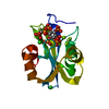

 PDBj
PDBj


 Assembly
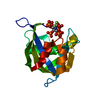


 Mass: 737.068 Da / Num. of mol.: 1 / Source method: obtained synthetically / Formula: C8H19F2NO24P6 / Feature type: SUBJECT OF INVESTIGATION
 Mass: 18.015 Da / Num. of mol.: 133 / Source method: isolated from a natural source / Formula: H2O
 Sample preparation
 Processing