 Movie
Movie Controller

[English] 日本語
 Yorodumi
- PDB-8g6i: Coagulation factor VIII bound to a patient-derived anti-C1 domain... -
+ Open data

- Basic information
| Entry | Database: PDB / ID: 8g6i | ||||||||||||||||||
|---|---|---|---|---|---|---|---|---|---|---|---|---|---|---|---|---|---|---|---|
| Title | Coagulation factor VIII bound to a patient-derived anti-C1 domain antibody inhibitor | ||||||||||||||||||
 Components Components |
| ||||||||||||||||||
 Keywords Keywords | BLOOD CLOTTING / Immune system | ||||||||||||||||||
| Function / homology |  Function and homology information Function and homology informationDefective F8 accelerates dissociation of the A2 domain / Defective F8 binding to the cell membrane / Defective F8 secretion / Defective F8 sulfation at Y1699 / Gamma carboxylation, hypusinylation, hydroxylation, and arylsulfatase activation / Defective F8 binding to von Willebrand factor / blood coagulation, intrinsic pathway / platelet alpha granule / Cargo concentration in the ER / Defective factor IX causes thrombophilia ...Defective F8 accelerates dissociation of the A2 domain / Defective F8 binding to the cell membrane / Defective F8 secretion / Defective F8 sulfation at Y1699 / Gamma carboxylation, hypusinylation, hydroxylation, and arylsulfatase activation / Defective F8 binding to von Willebrand factor / blood coagulation, intrinsic pathway / platelet alpha granule / Cargo concentration in the ER / Defective factor IX causes thrombophilia / Defective cofactor function of FVIIIa variant / Defective F9 variant does not activate FX / COPII-coated ER to Golgi transport vesicle / COPII-mediated vesicle transport / Defective F8 cleavage by thrombin / : / : / endoplasmic reticulum-Golgi intermediate compartment membrane / platelet alpha granule lumen / acute-phase response / Golgi lumen / blood coagulation / Platelet degranulation / oxidoreductase activity / endoplasmic reticulum lumen / copper ion binding / : / extracellular region / plasma membrane Similarity search - Function | ||||||||||||||||||
| Biological species |   Homo sapiens (human) Homo sapiens (human) | ||||||||||||||||||
| Method | ELECTRON MICROSCOPY / single particle reconstruction / cryo EM / Resolution: 4.23 Å | ||||||||||||||||||
 Authors Authors | Childers, K.C. / Davulcu, O. / Haynes, R.M. / Lollar, P. / Doering, C.B. / Coxon, C.H. / Spiegel, P.C. | ||||||||||||||||||
| Funding support |  United States, 5items United States, 5items
| ||||||||||||||||||
 Citation Citation | Journal: Blood / Year: 2023 Title: Structure of coagulation factor VIII bound to a patient-derived anti-C1 domain antibody inhibitor. Authors: Kenneth C Childers / Nathan G Avery / Kevin A Estrada Alamo / Omar Davulcu / Rose Marie Haynes / Pete Lollar / Christopher B Doering / Carmen H Coxon / P Clint Spiegel /  Abstract: The development of pathogenic antibody inhibitors against coagulation factor VIII (FVIII) occurs in ∼30% of patients with congenital hemophilia A receiving FVIII replacement therapy, as well as in ...The development of pathogenic antibody inhibitors against coagulation factor VIII (FVIII) occurs in ∼30% of patients with congenital hemophilia A receiving FVIII replacement therapy, as well as in all cases of acquired hemophilia A. KM33 is an anti-C1 domain antibody inhibitor previously isolated from a patient with severe hemophilia A. In addition to potently blocking FVIII binding to von Willebrand factor and phospholipid surfaces, KM33 disrupts FVIII binding to lipoprotein receptor-related protein 1 (LRP1), which drives FVIII hepatic clearance and antigen presentation in dendritic cells. Here, we report on the structure of FVIII bound to NB33, a recombinant derivative of KM33, via single-particle cryo-electron microscopy. Structural analysis revealed that the NB33 epitope localizes to the FVIII residues R2090-S2094 and I2158-R2159, which constitute membrane-binding loops in the C1 domain. Further analysis revealed that multiple FVIII lysine and arginine residues, previously shown to mediate binding to LRP1, dock onto an acidic cleft at the NB33 variable domain interface, thus blocking a putative LRP1 binding site. Together, these results demonstrate a novel mechanism of FVIII inhibition by a patient-derived antibody inhibitor and provide structural evidence for engineering FVIII with reduced LRP1-mediated clearance. | ||||||||||||||||||
| History |
|
- Structure visualization
| Structure viewer | Molecule: MolmilJmol/JSmol |
|---|
- Downloads & links
-Download
| PDBx/mmCIF format | 8g6i.cif.gz | 314.5 KB | Display | PDBx/mmCIF format |
|---|---|---|---|---|
| PDB format | pdb8g6i.ent.gz | 245.9 KB | Display | PDB format |
| PDBx/mmJSON format | 8g6i.json.gz | Tree view | PDBx/mmJSON format | |
| Others |  Other downloads Other downloads |
-Validation report
| Arichive directory | https://data.pdbj.org/pub/pdb/validation_reports/g6/8g6iftp://data.pdbj.org/pub/pdb/validation_reports/g6/8g6i | HTTPS FTP |
|---|
-Related structure data
| Related structure data |  29770MC M: map data used to model this data C: citing same article ( |
|---|---|
| Similar structure data |
-Links
 PDBj
PDBj


- Assembly
| Deposited unit | 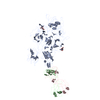
|
|---|---|
| 1 |
|
-Components
| #1: Protein | Mass: 168287.047 Da / Num. of mol.: 1 Source method: isolated from a genetically manipulated source Details: This protein is a chimera of human+pig coagulation factor VIII (PDB: 6MF0). Source: (gene. exp.) Homo sapiens (human)Gene: F8, CF8, F8, F8C / Production host:  Cricetulus griseus (Chinese hamster) / References: UniProt: P12263, UniProt: P00451 Cricetulus griseus (Chinese hamster) / References: UniProt: P12263, UniProt: P00451 | ||||
|---|---|---|---|---|---|
| #2: Antibody | Mass: 22972.436 Da / Num. of mol.: 1 Source method: isolated from a genetically manipulated source Source: (gene. exp.) Homo sapiens (human) / Production host: Cricetulus griseus (Chinese hamster) | ||||
| #3: Antibody | Mass: 22945.660 Da / Num. of mol.: 1 Source method: isolated from a genetically manipulated source Source: (gene. exp.) Homo sapiens (human) / Production host: Cricetulus griseus (Chinese hamster) | ||||
| #4: Polysaccharide | Source method: isolated from a genetically manipulated source Has ligand of interest | N | Has protein modification | Y | |
-Experimental details
-Experiment
| Experiment | Method: ELECTRON MICROSCOPY |
|---|---|
| EM experiment | Aggregation state: PARTICLE / 3D reconstruction method: single particle reconstruction |
- Sample preparation
| Component | Name: Structure of coagulation factor VIII bound to a patient-derived anti-C1 domain antibody inhibitor Type: COMPLEX Details: Coagulation factor VIII (molecule 1) is a chimera of human+pig proteins which was expressed in CHO cells. Entity ID: #1-#3 / Source: RECOMBINANT | |||||||||||||||
|---|---|---|---|---|---|---|---|---|---|---|---|---|---|---|---|---|
| Molecular weight | Value: 0.22 MDa / Experimental value: NO | |||||||||||||||
| Source (natural) | Organism: Homo sapiens (human) | |||||||||||||||
| Source (recombinant) | Organism: Cricetulus griseus (Chinese hamster) | |||||||||||||||
| Buffer solution | pH: 7.4 / Details: 150 mM NaCl, 20 mM HEPES pH 7.4 | |||||||||||||||
| Buffer component |
| |||||||||||||||
| Specimen | Conc.: 0.1 mg/ml / Embedding applied: NO / Shadowing applied: NO / Staining applied: NO / Vitrification applied: YES / Details: This sample was monodisperse. | |||||||||||||||
| Specimen support | Grid mesh size: 300 divisions/in. / Grid type: EMS Lacey Carbon | |||||||||||||||
| Vitrification | Instrument: FEI VITROBOT MARK I / Cryogen name: ETHANE / Humidity: 100 % / Chamber temperature: 277 K |
- Electron microscopy imaging
| Experimental equipment |  Model: Titan Krios / Image courtesy: FEI Company |
|---|---|
| Microscopy | Model: FEI TITAN KRIOS |
| Electron gun | Electron source:  FIELD EMISSION GUN / Accelerating voltage: 300 kV / Illumination mode: FLOOD BEAM FIELD EMISSION GUN / Accelerating voltage: 300 kV / Illumination mode: FLOOD BEAM |
| Electron lens | Mode: BRIGHT FIELD / Nominal defocus max: 2000 nm / Nominal defocus min: 800 nm / C2 aperture diameter: 100 µm |
| Image recording | Average exposure time: 1.565 sec. / Electron dose: 50 e/Å2 / Film or detector model: GATAN K3 (6k x 4k) / Num. of grids imaged: 1 / Num. of real images: 6993 |
- Processing
| EM software |
| ||||||||||||||||||||||||
|---|---|---|---|---|---|---|---|---|---|---|---|---|---|---|---|---|---|---|---|---|---|---|---|---|---|
| CTF correction | Type: PHASE FLIPPING AND AMPLITUDE CORRECTION | ||||||||||||||||||||||||
| Particle selection | Num. of particles selected: 8628762 | ||||||||||||||||||||||||
| Symmetry | Point symmetry: C1 (asymmetric) | ||||||||||||||||||||||||
| 3D reconstruction | Resolution: 4.23 Å / Resolution method: FSC 0.143 CUT-OFF / Num. of particles: 65133 / Symmetry type: POINT | ||||||||||||||||||||||||
| Atomic model building | PDB-ID: 6MF0 Accession code: 6MF0 / Source name: PDB / Type: experimental model |

