 Movie
Movie Controller

[English] 日本語
 Yorodumi
- PDB-8g6f: Structure of the Plasmodium falciparum 20S proteasome beta-6 A117... -
+ Open data

- Basic information
| Entry | Database: PDB / ID: 8g6f | ||||||
|---|---|---|---|---|---|---|---|
| Title | Structure of the Plasmodium falciparum 20S proteasome beta-6 A117D mutant complexed with inhibitor WLW-vs | ||||||
 Components Components |
| ||||||
 Keywords Keywords | HYDROLASE/HYDROLASE INHIBITOR / beta-6 A117D / inhibitor / proteasome / HYDROLASE / HYDROLASE-HYDROLASE INHIBITOR complex | ||||||
| Function / homology |  Function and homology information Function and homology informationER-Phagosome pathway / Cross-presentation of soluble exogenous antigens (endosomes) / Proteasome assembly / Antigen processing: Ub, ATP-independent proteasomal degradation / Orc1 removal from chromatin / CDK-mediated phosphorylation and removal of Cdc6 / FBXL7 down-regulates AURKA during mitotic entry and in early mitosis / KEAP1-NFE2L2 pathway / UCH proteinases / Ub-specific processing proteases ...ER-Phagosome pathway / Cross-presentation of soluble exogenous antigens (endosomes) / Proteasome assembly / Antigen processing: Ub, ATP-independent proteasomal degradation / Orc1 removal from chromatin / CDK-mediated phosphorylation and removal of Cdc6 / FBXL7 down-regulates AURKA during mitotic entry and in early mitosis / KEAP1-NFE2L2 pathway / UCH proteinases / Ub-specific processing proteases / Neddylation / Antigen processing: Ubiquitination & Proteasome degradation / MAPK6/MAPK4 signaling / AUF1 (hnRNP D0) binds and destabilizes mRNA / ABC-family protein mediated transport / Neutrophil degranulation / proteasome core complex / proteasome endopeptidase complex / proteasome core complex, beta-subunit complex / threonine-type endopeptidase activity / proteasome core complex, alpha-subunit complex / : / peptidase activity / endopeptidase activity / ubiquitin-dependent protein catabolic process / proteasome-mediated ubiquitin-dependent protein catabolic process / nucleus / cytoplasm / cytosol Similarity search - Function | ||||||
| Biological species |  | ||||||
| Method | ELECTRON MICROSCOPY / single particle reconstruction / cryo EM / Resolution: 2.58 Å | ||||||
 Authors Authors | Hsu, H.-C. / Li, H. | ||||||
| Funding support |  United States, 1items United States, 1items
| ||||||
 Citation Citation | Journal: Nat Commun / Year: 2023 Title: Structures revealing mechanisms of resistance and collateral sensitivity of Plasmodium falciparum to proteasome inhibitors. Authors: Hao-Chi Hsu / Daqiang Li / Wenhu Zhan / Jianxiang Ye / Yi Jing Liu / Annie Leung / Junling Qin / Benigno Crespo / Francisco-Javier Gamo / Hao Zhang / Liwang Cui / Alison Roth / Laura A ...Authors: Hao-Chi Hsu / Daqiang Li / Wenhu Zhan / Jianxiang Ye / Yi Jing Liu / Annie Leung / Junling Qin / Benigno Crespo / Francisco-Javier Gamo / Hao Zhang / Liwang Cui / Alison Roth / Laura A Kirkman / Huilin Li / Gang Lin /  Abstract: The proteasome of the malaria parasite Plasmodium falciparum (Pf20S) is an advantageous drug target because its inhibition kills P. falciparum in multiple stages of its life cycle and synergizes with ...The proteasome of the malaria parasite Plasmodium falciparum (Pf20S) is an advantageous drug target because its inhibition kills P. falciparum in multiple stages of its life cycle and synergizes with artemisinins. We recently developed a macrocyclic peptide, TDI-8304, that is highly selective for Pf20S over human proteasomes and is potent in vitro and in vivo against P. falciparum. A mutation in the Pf20S β6 subunit, A117D, confers resistance to TDI-8304, yet enhances both enzyme inhibition and anti-parasite activity of a tripeptide vinyl sulfone β2 inhibitor, WLW-vs. Here we present the high-resolution cryo-EM structures of Pf20S with TDI-8304, of human constitutive proteasome with TDI-8304, and of Pf20Sβ6 with WLW-vs that give insights into the species selectivity of TDI-8304, resistance to it, and the collateral sensitivity associated with resistance, including that TDI-8304 binds β2 and β5 in wild type Pf20S as well as WLW-vs binds β2 and β5 in Pf20Sβ6. We further show that TDI-8304 kills P. falciparum as quickly as chloroquine and artemisinin and is active against P. cynomolgi at the liver stage. This increases interest in using these structures to facilitate the development of Pf20S inhibitors that target multiple proteasome subunits and limit the emergence of resistance. | ||||||
| History |
|
- Structure visualization
| Structure viewer | Molecule: MolmilJmol/JSmol |
|---|
- Downloads & links
-Download
| PDBx/mmCIF format | 8g6f.cif.gz | 1.2 MB | Display | PDBx/mmCIF format |
|---|---|---|---|---|
| PDB format | pdb8g6f.ent.gz | 1 MB | Display | PDB format |
| PDBx/mmJSON format | 8g6f.json.gz | Tree view | PDBx/mmJSON format | |
| Others |  Other downloads Other downloads |
-Validation report
| Arichive directory | https://data.pdbj.org/pub/pdb/validation_reports/g6/8g6fftp://data.pdbj.org/pub/pdb/validation_reports/g6/8g6f | HTTPS FTP |
|---|
-Related structure data
| Related structure data |  29765MC 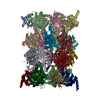 8g6eC  8ud9C M: map data used to model this data C: citing same article ( |
|---|---|
| Similar structure data |
-Links
 PDBj
PDBj


- Assembly
| Deposited unit | 
|
|---|---|
| 1 |
|
-Components
-Proteasome subunit alpha type- ... , 7 types, 14 molecules AOBPCQDRESFTGU
| #1: Protein | Mass: 29531.656 Da / Num. of mol.: 2 / Source method: isolated from a natural source / Source: (natural) References: UniProt: Q8IAR3, proteasome endopeptidase complex #2: Protein | Mass: 26556.391 Da / Num. of mol.: 2 / Source method: isolated from a natural source / Source: (natural) #3: Protein | Mass: 27977.664 Da / Num. of mol.: 2 / Source method: isolated from a natural source / Source: (natural) #4: Protein | Mass: 27263.285 Da / Num. of mol.: 2 / Source method: isolated from a natural source / Source: (natural) #5: Protein | Mass: 28417.367 Da / Num. of mol.: 2 / Source method: isolated from a natural source / Source: (natural) #6: Protein | Mass: 28871.697 Da / Num. of mol.: 2 / Source method: isolated from a natural source / Source: (natural) #7: Protein | Mass: 29324.295 Da / Num. of mol.: 2 / Source method: isolated from a natural source / Source: (natural) References: UniProt: O77396, proteasome endopeptidase complex |
|---|
-Protein , 1 types, 2 molecules HV
| #8: Protein | Mass: 29143.936 Da / Num. of mol.: 2 / Source method: isolated from a natural source / Source: (natural) |
|---|
-Proteasome subunit beta- ... , 6 types, 12 molecules IWJXKYLZMaNb
| #9: Protein | Mass: 25104.885 Da / Num. of mol.: 2 / Source method: isolated from a natural source / Source: (natural) #10: Protein | Mass: 24533.131 Da / Num. of mol.: 2 / Source method: isolated from a natural source / Source: (natural) #11: Protein | Mass: 22889.105 Da / Num. of mol.: 2 / Source method: isolated from a natural source / Source: (natural) #12: Protein | Mass: 23620.646 Da / Num. of mol.: 2 / Source method: isolated from a natural source / Source: (natural) #13: Protein | Mass: 27345.213 Da / Num. of mol.: 2 / Mutation: A117D / Source method: isolated from a natural source / Source: (natural) #14: Protein | Mass: 30909.893 Da / Num. of mol.: 2 / Source method: isolated from a natural source / Source: (natural) |
|---|
-Non-polymers , 2 types, 603 molecules 

| #15: Chemical | ChemComp-7F1 / (  Mass: 690.852 Da / Num. of mol.: 4 / Source method: obtained synthetically / Formula: C36H46N6O6S / Feature type: SUBJECT OF INVESTIGATION Mass: 690.852 Da / Num. of mol.: 4 / Source method: obtained synthetically / Formula: C36H46N6O6S / Feature type: SUBJECT OF INVESTIGATION#16: Water | ChemComp-HOH / | Mass: 18.015 Da / Num. of mol.: 599 / Source method: isolated from a natural source / Formula: H2O |
|---|
-Details
| Has ligand of interest | Y |
|---|---|
| Has protein modification | Y |
-Experimental details
-Experiment
| Experiment | Method: ELECTRON MICROSCOPY |
|---|---|
| EM experiment | Aggregation state: PARTICLE / 3D reconstruction method: single particle reconstruction |
- Sample preparation
| Component | Name: the Plasmodium falciparum 20S proteasome beta-6 A117D mutant complexed with inhibitor WLW-vs Type: COMPLEX / Entity ID: #1-#14 / Source: NATURAL | ||||||||||||||||||||||||||||||
|---|---|---|---|---|---|---|---|---|---|---|---|---|---|---|---|---|---|---|---|---|---|---|---|---|---|---|---|---|---|---|---|
| Molecular weight | Value: 0.7 MDa / Experimental value: YES | ||||||||||||||||||||||||||||||
| Source (natural) | Organism: | ||||||||||||||||||||||||||||||
| Buffer solution | pH: 7.5 | ||||||||||||||||||||||||||||||
| Buffer component |
| ||||||||||||||||||||||||||||||
| Specimen | Conc.: 0.5 mg/ml / Embedding applied: NO / Shadowing applied: NO / Staining applied: NO / Vitrification applied: YES / Details: partially purified | ||||||||||||||||||||||||||||||
| Specimen support | Grid material: GOLD / Grid mesh size: 300 divisions/in. / Grid type: Quantifoil R1.2/1.3 | ||||||||||||||||||||||||||||||
| Vitrification | Instrument: FEI VITROBOT MARK IV / Cryogen name: ETHANE / Humidity: 100 % / Chamber temperature: 279 K |
- Electron microscopy imaging
| Experimental equipment |  Model: Titan Krios / Image courtesy: FEI Company |
|---|---|
| Microscopy | Model: FEI TITAN KRIOS |
| Electron gun | Electron source:  FIELD EMISSION GUN / Accelerating voltage: 300 kV / Illumination mode: FLOOD BEAM FIELD EMISSION GUN / Accelerating voltage: 300 kV / Illumination mode: FLOOD BEAM |
| Electron lens | Mode: BRIGHT FIELD / Nominal magnification: 105000 X / Nominal defocus max: 1800 nm / Nominal defocus min: 1300 nm / Cs: 2.7 mm / C2 aperture diameter: 100 µm / Alignment procedure: COMA FREE |
| Specimen holder | Cryogen: NITROGEN / Specimen holder model: FEI TITAN KRIOS AUTOGRID HOLDER |
| Image recording | Average exposure time: 1.3 sec. / Electron dose: 60 e/Å2 / Film or detector model: GATAN K3 BIOQUANTUM (6k x 4k) / Num. of grids imaged: 1 / Num. of real images: 24806 |
| EM imaging optics | Energyfilter name: GIF Bioquantum / Energyfilter slit width: 20 eV |
| Image scans | Width: 11520 / Height: 8184 |
- Processing
| EM software |
| ||||||||||||||||||||||||||||||||||||||||
|---|---|---|---|---|---|---|---|---|---|---|---|---|---|---|---|---|---|---|---|---|---|---|---|---|---|---|---|---|---|---|---|---|---|---|---|---|---|---|---|---|---|
| CTF correction | Type: PHASE FLIPPING AND AMPLITUDE CORRECTION | ||||||||||||||||||||||||||||||||||||||||
| Particle selection | Num. of particles selected: 337322 | ||||||||||||||||||||||||||||||||||||||||
| Symmetry | Point symmetry: C2 (2 fold cyclic) | ||||||||||||||||||||||||||||||||||||||||
| 3D reconstruction | Resolution: 2.58 Å / Resolution method: FSC 0.143 CUT-OFF / Num. of particles: 74883 / Algorithm: BACK PROJECTION / Num. of class averages: 3 / Symmetry type: POINT | ||||||||||||||||||||||||||||||||||||||||
| Atomic model building | B value: 43.89 / Protocol: RIGID BODY FIT / Space: REAL | ||||||||||||||||||||||||||||||||||||||||
| Atomic model building | PDB-ID: 8G6E Accession code: 8G6E / Source name: PDB / Type: experimental model | ||||||||||||||||||||||||||||||||||||||||
| Refine LS restraints |
|
